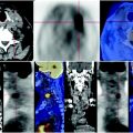
Fig. 71.1
CT shows dysmorphic kidneys, sharply increased in size, structure subverted by numerous coarse parenchymal cysts that have abnormal deposition of the tracer at the PET scan. Absence of focal lesions
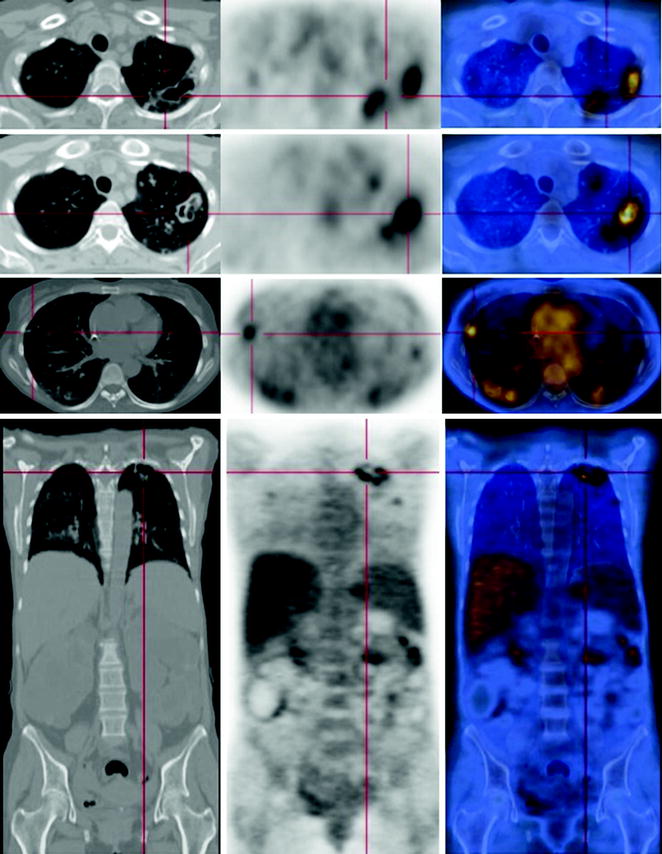
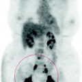
Fig. 71.2
At the apical segment of the left upper lobe, CT-PET image shows an oval lesion with air inside, with unevenly thickened margins that express low metabolic activity. Multiple bilateral pulmonary nodules of various sizes, some measuring more than a centimeter, other millimetric, can be seen
Related posts:
 Laryngeal Squamous Carcinoma: Staging
 Radio-Treated Cancer of the Posterior Hemi-Circumference of the Anal Canal: Post-Actinic Fibrosis
 Bone-Destroying Metastases in Thyroid Undifferentiated Carcinoma
 Surgically Treated Endometrial Cancer: Bilateral Nodal Recurrence
 Undifferentiated Gastric Adenocarcinoma with Peritoneal Carcinosis
 Follow-Up of Papillary Breast Cancer: Solitary Sacroiliac Benign Lesion
Stay updated, free articles. Join our Telegram channel

Full access? Get Clinical Tree


