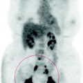
Fig. 72.1
The PET scan shows high metabolic activity secondary to post actinic reaction at the pulmonary hilum and right basal pleural granulomatous reaction secondary to ipsilateral talcage
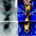
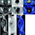
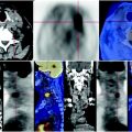
Increased FDG deposition in multiple nodules at the parietal, mediastinal and diaphragmatic right pleura, SUV max 4.
Absent glucose consumption of the renal right lower polar and ipsilateral adrenal mass. No focal areas of abnormal metabolism in the remaining parts of the body examined.
72.4 Conclusions
The PET scan shows high metabolic activity secondary to post actinic reaction at the pulmonary hilum and right basal pleural granulomatous reaction secondary to ipsilateral talcage (Figs. 72.1, 72.2).
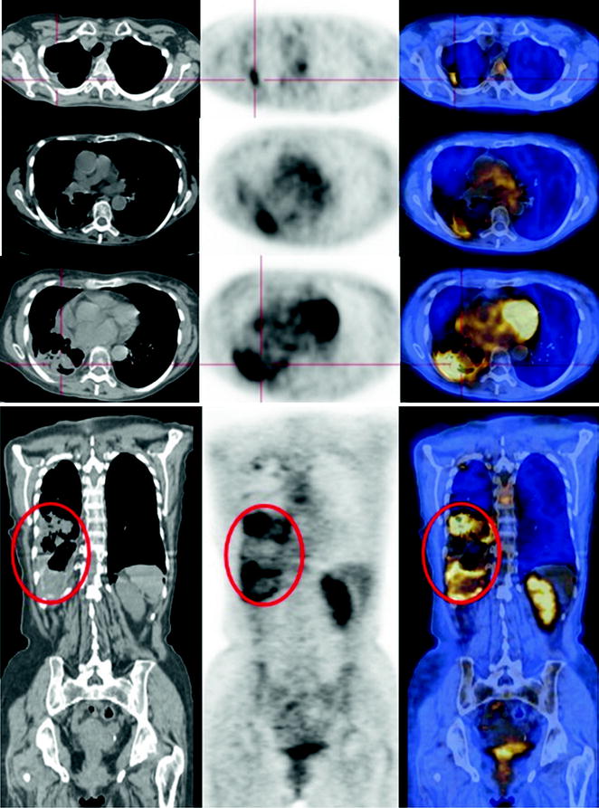
Fig. 72.2
At the posterior basal segment of the right lower lobe, a CT scan shows a large area of consolidation tissue that PET shows to have limited consumption of glucose and is therefore to be correlated with actinic outcomes. There can also be seen multiple pleural high FDG metabolism lumps determined by the granulomatous reaction to the talcage
72.5 Key Points
In the treatment for lung cancer, radiotherapy determines an actinic reaction, sometimes so intense that decreases substantially only over time. The volume of lung irradiated, which is expected to be limited (as small as possible), the total administered dose, fractionation, and the combination with chemotherapy and concomitant respiratory diseases are all risk factors that contribute to a stabilized parenchymal damage: actinic fibrosis.
Related posts:
 Laryngeal Squamous Carcinoma: Staging
 Radio-Treated Cancer of the Posterior Hemi-Circumference of the Anal Canal: Post-Actinic Fibrosis
 Bone-Destroying Metastases in Thyroid Undifferentiated Carcinoma
 Surgically Treated Endometrial Cancer: Bilateral Nodal Recurrence
 Undifferentiated Gastric Adenocarcinoma with Peritoneal Carcinosis
 Follow-Up of Papillary Breast Cancer: Solitary Sacroiliac Benign Lesion
Stay updated, free articles. Join our Telegram channel

Full access? Get Clinical Tree


