Pulmonary Edema (Interstitial)
Sam A. Glaubiger
CLINICAL HISTORY
58-year-old male with shortness of breath.
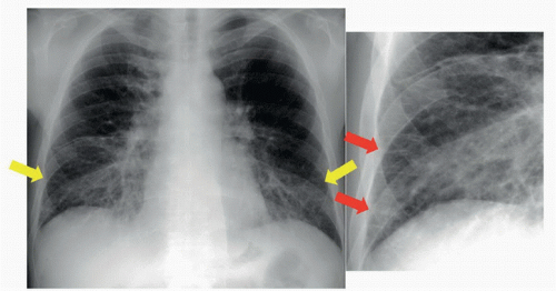 FIGURE 70A |
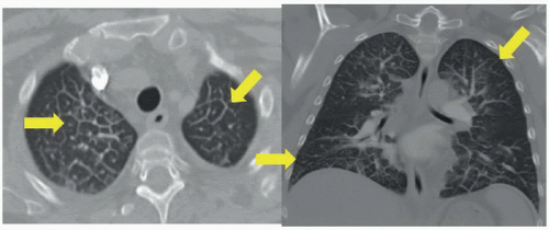 FIGURE 70B |
FINDINGS
Figure 70A: Posteroanterior (PA) plain film of the chest and coned-down view of the same image. Kerley B lines, or thickened interlobular septa (yellow arrows). Coned-down view shows Kerley B lines extending to the pleura (red arrows). The hila are indistinct and assume a “fluffy” appearance. Figure 70B: Axial and coronal CT image of the chest. Both images show interlobular septal thickening (yellow arrows), outlining the secondary pulmonary lobule.
DIFFERENTIAL DIAGNOSIS
Pulmonary edema (interstitial), pulmonary fibrosis, lymphangitic carinomatosis, sarcoidosis.
Related posts:


Stay updated, free articles. Join our Telegram channel

Full access? Get Clinical Tree


