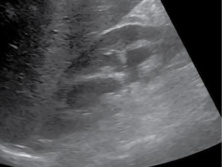
Pyonephrosis is accumulation of pus in an obstructed pelvicalyceal system with suppurative destruction of the renal parenchyma. It is a medical emergency and early recognition of this condition is critical, as emergent decompression is needed to prevent rapid decline in renal function. In a patient with known obstructive uropathy, fever and flank plain should raise a red flag for possible pyonephrosis. There are a variety of causes of urinary obstruction (summarized in Fig. 10.12.2.4.9) and may be due to ureteric calculi, stricture, developmental ureteric anomalies, retroperitoneal fibrosis or malignant disease. End-stage chronic pyelonephritis may also result in pyonephrotic kidney. Plain X-ray abdomen may show enlarged kidney with or without stones. Intravenous Urogram shows poorly or nonfunctioning kidney. It is often the first diagnostic imaging study, demonstrating dilated collecting system with dependent echoes and shifting debris representing purulent inflammatory contents, which helps to distinguish this condition from simple hydronephrosis (Fig. 10.12.2.4.10). The echogenic debris may appear mass like, thus confusing with haemorrhage or tumour; however avascularity on Doppler interrogation suggests pyonephrosis.
Pyonephrosis
Introduction
Aetiology

Imaging findings
Plain X-ray abdomen
Intravenous urogram
Ultrasound
Related posts:


Stay updated, free articles. Join our Telegram channel

Full access? Get Clinical Tree


