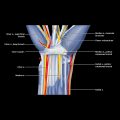
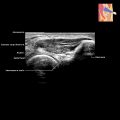
Radial Nerve TERMINOLOGY Abbreviations • Extensor indicis (EI) GROSS ANATOMY Arm • Radial nerve is largest branch of posterior cord of brachial plexus Receives contributions from cervical roots C5-C8 Contains motor and sensory components that supply extensor muscles of arm and forearm Lies posterior and deep to axillary artery in axilla • Lies between coracobrachialis and teres major muscles and then between bellies of medial and lateral head of triceps • Runs deep to triceps in spiral groove and then deep to brachioradialis • 10 cm proximal to lateral epicondyle of humerus, radial nerve penetrates lateral intermuscular septum to enter anterior compartment of arm • Area between lateral intermuscular septum and supinator inlet known as radial tunnel • Just anterior to lateral epicondyle, nerve bifurcates into sensory (superficial) and motor (deep) branches Forearm • Branches of radial nerve Superficial branch of radial nerve (purely sensory) – Courses distally, deep to brachioradialis, to extensor compartment of distal forearm and then to dorsal wrist – Cephalic vein crosses over superficial branch in distal forearm Nerve may be injured during venous cannulation – Divides into lateral and medial branches 5 cm proximal to radial styloid – Lateral branch supplies radial wrist and thumb skin; medial branch (supplies mid and ulnar wrist skin) Dorsal digital nerves supply ulnar thumb, index, middle, and radial ring fingers Deep branch of radial nerve (purely motor) – Deep branch of radial nerve becomes PIN at entry into supinator muscle (supinator arch or arcade of Fröhse) – PIN passes between superficial and deep heads of supinator muscle (supinator canal) Then exits distally and runs posteriorly as PIN – Supplies extensor compartment muscles of forearm – 5 potential anatomical causes of PIN compression near elbow Fibrous band of radial head, which is continuous with extensor carpi radialis brevis (ECRBr) and superficial head of supinator muscle Radial recurrent vessels (leash of Henry) at level of radial head Tendinous margin of ECRBr Aponeurotic proximal margin of supinator (arcade of Fröhse) = most common site of compression Distal margin of supinator muscle – After exiting from supinator canal, PIN travels between superficial and deep extensor muscles until it reaches interosseous membrane distally Travels 1st with posterior interosseous artery and then anterior interosseous artery – At wrist, PIN lies deep to radial side of 4th extensor compartments where it can be focally enlarged (pseudoganglion) Thereafter, PIN divides into 2-4 terminal branches Hand: Branches of Radial Nerve • Not usually seen on routine imaging • No motor innervation in hand • Sensory to back of hand and dorsal aspect of radial 3.5 digits ANATOMY IMAGING ISSUES Imaging Recommendations • Radial nerve is best identified at lateral aspect of distal arm deep to brachioradialis muscle Alternatively, identify nerve deep to triceps in spiral groove midhumeral level Or in proximal aspect of arm medially – Lying just deep to axillary and brachial arteries Only gold members can continue reading. Log In or Register to continue Share this:Click to share on Twitter (Opens in new window)Click to share on Facebook (Opens in new window) Related posts: Hand Vessels Elbow Soft Tissue Tumor Biopsy Hypoechoic Muscle Mass Hip and Pelvis Procedures Baker Cyst Stay updated, free articles. Join our Telegram channel Join Tags: Diagnostic Ultrasound Musculoskeletal Apr 9, 2020 | Posted by admin in MUSCULOSKELETAL IMAGING | Comments Off on Radial Nerve Full access? Get Clinical Tree