CHAPTER 10 Radiation Dose Reduction Strategies in Cardiac Computed Tomography
Rapid technical advances in CT and increased availability of cardiac-capable CT systems have led to a sharp increase in the number of cardiac CT examinations performed during the last decade. Some technical advances (smaller detectors, faster gantry rotation) have necessitated increased x-ray exposure to the patient to maintain image noise. Concern regarding the resulting increase in radiation dose to the population from cardiac CT and the associated biologic risk has motivated critical assessment of dose-related imaging parameters.
A strategy for reducing radiation dose to the patient undergoing cardiac imaging should be employed particularly for patients at greatest risk for harm from x-ray exposure to the chest, young patients, and female patients.1 This plan should include educating patients on the risks of exposure to ionizing radiation, seeking alternative studies that do not rely on ionizing radiation (e.g., MRI or ultrasonography) when appropriate, assessing the risk/benefit ratio of CT for the individual patient, and applying the as low as reasonably achievable (ALARA) principle to the selection of CT scan parameters.
TECHNICAL REQUIREMENTS
This chapter focuses on the selection of critical scan parameters directly or indirectly affecting radiation dose. Numerous modifiable scan parameters, including x-ray tube current, peak x-ray tube voltage, pitch (for helical scanning) or image acquisition spacing (for axial scanning), scan length, and, on some systems, scan field of view [SFOV], directly influence radiation dose by altering the number or energy of the x-ray photons interacting with the patient. Other scan parameters, including reconstructed slice thickness, indirectly affect dose by changing image quality and prompting the scanner operator to change parameters directly influencing dose. Strategies for customizing these parameters for the individual patient and minimizing radiation dose are presented in general terms to benefit users of any CT system.
TECHNIQUE TO REDUCE X-RAY TUBE CURRENT
ECG-Based Reduction in Tube Current
Acquisition of data using a lower dose, axial mode is indicated for contrast-enhanced evaluation of the coronary arteries in patients with low, stable heart rates; non–contrast-enhanced evaluation of coronary artery calcium (also known as calcium scoring), and evaluation of less mobile cardiovascular anatomy (e.g., aorta) if the z coverage of the scanner allows a reasonable breath-hold time. Helical data acquisition is typically required for contrast-enhanced coronary evaluation in patients with high or irregular heart rates. Online, ECG-based modulation of the tube current should be used with helical techniques to the extent possible for a given scanner.
Size-Based Reduction in Tube Current
Reduction of the nominal x-ray tube current before scanning is indicated in thin patients imaged with axial or helical techniques to reduce radiation dose. Online, anatomic-based tube current modulation during scanning can be used for all patients imaged with axial and non–ECG-gated, helical techniques.
Technique Description
X-rays used for CT imaging are generated when accelerated electrons strike a tungsten target. The number of electrons striking the target per unit time is described as the x-ray tube current and expressed in units of milliamperes (mA). A decrease in the x-ray tube current decreases the number of electrons striking the target and, subsequently, the number of x-rays produced per unit time.
Radiation dose decreases linearly with a decrease in tube current such that a 20% reduction in tube current results in a 20% reduction in dose. Dose savings is achieved at the expense, however, of increased image noise because decreasing the number of x-ray photons produced per unit time decreases the probable number of photons penetrating the patient and reaching the detector array. Image noise is proportional to such that a 20% reduction in tube current results in a 12% increase in image noise.
The parameter manipulated on clinically available CT scanners is often the product of the tube current and the exposure time per rotation with units of milliampere-second (mAs). The tube current × time product determines the number of x-ray photons produced per rotation. Additionally, some manufacturers automatically normalize the tube current × time product to pitch for helical scanning and define the resulting value in units of effective mAs or mAs/slice.
ECG-Based Reduction in Tube Current
During cardiac imaging, the x-ray tube current can be switched off or greatly reduced during systolic phases of the cardiac cycle significantly decreasing radiation dose. In patients with low, stable heart rates, data acquisition can be confined to a limited portion of the RR interval using prospectively ECG-triggered axial techniques. Average effective radiation doses of 2 to 3 mSv have been reported using these techniques to image the coronary arteries.2,3
Axial imaging is associated with increased sensitivity to arrhythmia, owing to prospective referencing of the ECG signal, and increased examination times, owing to incrementing the patient table between acquisitions. It is often necessary to acquire data during the entire cardiac cycle using a helical mode and retrospectively reference image reconstruction to a simultaneously recorded ECG signal. Although ECG-gated helical techniques require continuous x-ray exposure, tube current outside the phase of interest can be reduced to decrease patient radiation dose significantly. Effective radiation doses can easily exceed 15 mSv4 using helical imaging without ECG-based dose modulation, but can be reduced 50% or more depending on patient heart rate,5 the minimum tube current value,6 and the duration of the full tube current window.7
Size-Based Reduction in Tube Current
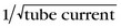
The x-ray tube current can be reduced for slimmer patients. Attenuation of the incident x-ray beam decreases with the thickness of the tissue between the x-ray source and the detector such that less radiation exposure is required to penetrate thinner tissues and achieve desired image noise (Fig. 10-1). Patients can be assigned to size categories based on visual inspection, weight, body mass index, or cross-sectional body measurements from scout images,8 and the tube current can be adjusted manually to a predefined value. Weight-adapted tube current protocols were shown to reduce coronary CT angiography dose by 18% in men and 26% in women9 at one institution.

FIGURE 10-1 A and B, Axial images from a larger patient (lateral width at top of liver 40 cm) (A) and a smaller patient (lateral width 27 cm) (B). To achieve noise equal to approximately 25 HU, 400 mAs/rotation was required in the larger patient, whereas only 320 mAs/rotation was required to achieve the same noise level in the smaller patient. Data acquisition with a lower tube current and resultant decrease in patient x-ray exposure is acceptable for imaging of smaller compared with larger patients without increasing image noise.
Automatic methods of online adaptation of tube current to patient size can also be used to reduce dose. The tube current can be modulated along the x, y, and z directions during scanning based on local tissue thickness without sacrificing image noise. Tube current is reduced at projection angles and table positions requiring less x-ray penetration. Online, anatomic-based tube current modulation has been shown to reduce radiation exposure to the thorax by 20% compared with a fixed tube current while maintaining image noise.10
Pitfalls and Solutions
ECG-Based Reduction in Tube Current
Axial imaging is vulnerable to cardiac motion artifacts, particularly in patients with high or irregular heart rates, according to prospective data collection. Some additional data beyond the minimum required for image reconstruction can be acquired (also known as padding) to permit minor retrospective adjustments of the reconstruction window and, potentially, to reduce cardiac motion artifacts.
Helical data acquisition with retrospective ECG gating is less susceptible to cardiac motion artifacts and provides an alternative to axial imaging at high or irregular heart rates. ECG-based tube current modulation is prescribed before scanning, however, so changes in heart rate could result in unintended reduction of the tube current during a desired phase of reconstruction for a given cardiac cycle. Some CT systems allow adjustment of the full tube current duration, increasing the utility of ECG-based tube current modulation for patients with high or irregular heart rates because the optimal reconstruction phase is less predictable.7

Stay updated, free articles. Join our Telegram channel

Full access? Get Clinical Tree

