Radiation source
Energy
Radium-226
830 keV
Caesium-137
662 keV
Iridium-192
380 keV
Electron linacs
4–12 MeV
INTRABEAM
50 kV
Intraoperative radiotherapy has many benefits, as described in previous chapters, and can be performed using electron linear accelerators (linacs, e.g. Mobetron, IntraOp Medical Inc and Liac, Sordina SpA) operating at megavoltage energies. More recent designs of these units are self-shielding with beam stoppers, but there may still be limitations to the number of procedures that can be performed (Daves and Mills 2001; Soriani et al. 2010).
The INTRABEAM system operates at a peak voltage of 50 kV, so has a much lower energy than afterloading brachytherapy or electron linac approaches. This greatly simplifies the shielding requirements, yet maintains the benefits of short range delivery with rapid attenuation of dose, an electronic source that can be readily switched on and off, and the efficiency of treatment during surgery. These combined features have led to the title of “electronic brachytherapy” (Park et al. 2010). Caution is still advised, since the unshielded dose rate from the source can reach 1 Gy/min, much higher than a diagnostic X-ray source with which theatre staff may be familiar. However, shielding requirements are practical and straightforward, as will be described in this chapter.
5.2 Legislation
Hazards associated with higher doses of radiation have led to strict legal requirements, which will be specific to each country. Most of Europe derives its laws from the European Union directives, given in Table 5.2. There will also usually be a national license or authorisation to use radiation, which must be obtained before beginning to use any material or equipment.
Table 5.2
Summary of legal requirements in Europe
Directive 96/29/Euratom |
Consultation with Qualified (radiation) Expert: |
Controlled area definition and working instructions |
Prior critical examination and acceptance |
Regular checking and calibration |
Dose monitoring and training |
Directive 97/43/Euratom |
Justification, optimisation and limitation of exposures |
Medical Physics Expert closely involved in radiotherapeutic practices |
In the UK, safety of staff and members of the public is covered by the Ionising Radiation Regulations (1999) of the Health and Safety at Work Act (1974). These define the role of a Radiation Protection Adviser, who is the qualified radiation expert for the organisation, and a Radiation Protection Supervisor, who supervises compliance with the specific procedures relating to the safety of the radiation technique (the Local Rules). Safety of the patient is covered by the Ionising Radiation (Medical Exposure) Regulations (2000), which require any radiation exposure to be justified by a defined practitioner (usually a radiation oncologist) and delivered by defined operators. Both the staff operating the INTRABEAM system and the surgeon who positions the applicator are considered to be operators and should be named in the local procedures.
In the USA, the system is still considered to be an emerging technology and although it has received Food and Drugs Administration clearance, it is not currently covered by Nuclear Regulatory Commission regulations. The specific requirements of each state will also vary. Guidelines have been produced by the American Association of Physicists in Medicine (AAPM, Thomadsen et al. 2009), which are summarised in Table 5.3.
Table 5.3
Summary of legal requirements in the USA
AAPM guidelines |
|---|
The services of an Authorised Medical Physicist shall be required in facilities using electronic brachytherapy units: |
Dosimetry and treatment calculations |
Calibration validation and quality assurance |
Shielding and area designation |
Dose monitoring and training Physically present during treatments |
All of these documents describe the close involvement of experts in radiation physics, who should be consulted with regard to both commissioning a new service and ongoing clinical support.
5.3 Basics of Radiation Protection
The philosophical basis underlying radiation safety is to keep doses to staff (and others not undergoing treatment) as low as reasonably practicable (ALARP).2
Four general principles can be used to achieve this:
Time
Distance
Shielding
Contamination
Radiation exposure is directly proportional to time, but follows an inverse square dependence on distance. For example, if the distance is doubled, the exposure is reduced by a factor of 4. Therefore distance can have a large impact on doses received. As mentioned earlier, less shielding is required for lower energy X-rays.
Because the INTRABEAM system is an electronic X-ray source, there is no issue of contamination, since the radiation exposure only occurs when the beam is on. If any intervention for the patient is required, the radiation can be switched off (paused) and poses no further hazard during this time.
Radiation dose may be measured using a number of different units. The most fundamental is the Gray (Gy), which is commonly used to describe the amount of radiation given therapeutically, since it corresponds to the energy deposited per unit mass and can be readily measured. However, effective doses to individuals for protection considerations are quoted in Sieverts (Sv), which take account of the relative biological effect of different types of radiation and different sensitivities of different tissues in the body. Typical values are thousandths (mSv) or millionths (μSv) of this unit, and the latter will be used for all subsequent quoted values, since many detectors read in this scale.
Although current models for risk from radiation equate any dose with some risk, it is important to consider small exposures in context. Every individual receives some exposure to ionising radiation from natural background sources, listed in Table 5.4, as well as any medical exposures. Some comparative doses for common activities, legislative limits and adverse effects are given in Table 5.5.
Table 5.4
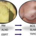 Targeted Intraoperative Radiotherapy: Concept and Review of Evidence in Breast Cancer
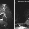 Follow-Up Findings in the Tumour Bed After IORT – What the Radiologist Needs to Know
 Quality Assurance and Training of Targeted Intraoperative Radiotherapy: Establishment of the TARGIT Academy
 Treating Patients with TARGIT
 Intraoperative Photon Radiosurgery in Patients with Malignant Brain Tumours
 Radiobiology


Sources of annual (background) radiation doses to the UK population
Related posts:
Targeted Intraoperative Radiotherapy: Concept and Review of Evidence in Breast Cancer
Follow-Up Findings in the Tumour Bed After IORT – What the Radiologist Needs to Know
Quality Assurance and Training of Targeted Intraoperative Radiotherapy: Establishment of the TARGIT Academy
Treating Patients with TARGIT
Intraoperative Photon Radiosurgery in Patients with Malignant Brain Tumours
Radiobiology
Stay updated, free articles. Join our Telegram channel
Full access? Get Clinical Tree
