Radio-Occult Hip Fracture
Ami V. Vakharia
Daniel B. Nissman
CLINICAL HISTORY
76-year-old woman fell on her left side while walking at the supermarket and now complains of hip pain. Radiographs are negative for fracture.
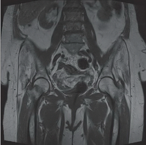 FIGURE 80A |
 FIGURE 80B |
FINDINGS
Coronal T1-weighted MR image (Fig. 80A) through the entire pelvis reveals a curvilinear hypointensity within the lateral right subcapital femoral neck that extends approximately half the width of the femoral neck with adjacent, more ill-defined hypointensity consistent with edema. Coronal T2-weighted fat-suppressed image (Fig. 80B) of only the right hip demonstrates a curvilinear hypointensity that matches the course of the hypointensity on the T1-weighted image with a thin rim of marked adjacent hyperintensity. More ill-defined hyperintensity of the adjacent bone marrow is also noted. Radiographs immediately preceding the MRI were normal.
DIFFERENTIAL DIAGNOSIS
Osteoarthrosis, intertrochanteric bursitis, radio-occult hip fracture, soft tissue tumor or metastases.
DIAGNOSIS
Radio-occult right subcapital femoral neck fracture.
DISCUSSION
Hip fractures in the elderly are associated with high morbidity and 1-year mortality, which may be as high as 30%.1 A nondisplaced hip fracture, particularly in the elderly patient with osteopenia, is at times difficult to visualize. With our population increasingly outliving historical average age estimates, hip fractures are expected to double in the next 20 years. The typical mechanism in the elderly is minor trauma, usually a fall from a standing position, to a bone that is already weakened by osteoporosis.
Related posts:


Stay updated, free articles. Join our Telegram channel

Full access? Get Clinical Tree


