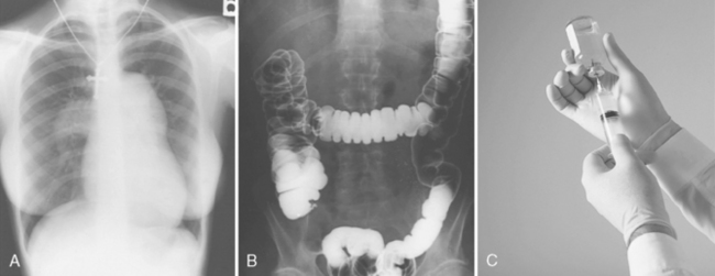
CHAPTER 9 On completion of this chapter, you should be able to: • Describe internal and external patient preparation. • Describe examinations that use iodine, barium, or air as a contrast agent. • Describe what is included in skull and headwork radiography. • Describe what is included in thoracic radiography. • Describe what is included in extremity radiography. • Describe what is included in spinal radiography. • List conditions for which abdominal radiography may be performed. • Briefly explain esophagograms, upper gastrointestinal series, small bowel studies, and barium enemas. • List and describe the examinations that are referred to as special procedures. Depending on the examination, patient preparation is performed either internally or externally. External preparation requires removing clothing and jewelry that may be covering the area of the body through which the x-rays must pass. Many types of clothing material show up on film as obscure shadows. Buttons and zippers may hide small disease processes or fractures. If a region of the head is being radiographed, dentures must be removed because they interfere with the passage of x-rays through the mouth. Rings and watches must be removed when radiographing the hand or wrist. One of the most common mistakes is forgetting to remove a necklace before performing a chest examination (Fig. 9-1, A). Always ask each patient to remove jewelry before beginning an examination. In addition, ask the patient if the area of the body to be radiographed is pierced; if so, request that all piercing jewelry be removed from the body part. Failure to do so results in a double dose of radiation to the patient because the radiographs must be retaken. Note if a tattoo is in the area being radiographed. If the ink used in the tattoo contains metallic pigmentation, it may appear as a faint shadow on the radiographic image. Proper examination of the patient is the responsibility of the radiographer. Checks for unwanted objects should be verbal, visual, and tactile. The element barium has approximately the same contrast qualities as iodine, but the similarities end there. Barium sulfate is inert and cannot be absorbed by the body; this makes it the medium of choice for gastrointestinal studies (Fig. 9-1, B-C). Patient allergic reaction to barium is almost nonexistent because of its inert properties.
Radiographic Examinations
Diagnosing Disease and Injury
Patient preparation and contrast media
![]()
Stay updated, free articles. Join our Telegram channel

Full access? Get Clinical Tree


Radiology Key
Fastest Radiology Insight Engine