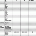
Depth of invasion (No. of patients)
Series
Primary site
≤1 mm
1.01–2.00 mm
2.01–4.00 mm
>4.00 mm
Rousseau et al. [39], M.D. Anderson Hospital
Various
4 % (388)
12 % (522)
28 % (314)
44 % (151)
Emery et al. [40], University of Oregon
Various
2 % (41)
13 % (85)
20 % (35)
27 % (11)
Paek et al. [41], University of Michigan
Various
–
19 % (490)
32 % (301)
45 % (119)
Kruper et al. [42], University of Pennsylvania
Various
5 % (251)
10 % (228)
20 % (140)
38 % (63)
Leong et al. [43], Multicenter
Head and neck
3 % (134)
7 % (230)
21 % (160)
13 % (63)
Berk et al. [44], Stanford University
Various
0 % (45)
18 % (115)
19 % (64)
16 % (32)
The likelihood of nodal recurrence in a negative SLNB basin is relatively low. Vuylsteke and colleagues reported on 209 patients with stage I and II melanomas who underwent SLNB at the Vrije Universiteit between 1993 and 1996 [36]. SLNB was successful in 208 of 209 patients (99 %) and survivors were followed for 5 years. Four of 168 patients (2 %) with a negative SLNB recurred in the nodal basin and 11 patients (7 %) developed a local-regional dermal recurrence.
Incidence of Residual Positive Nodes in a Completion Node Dissection After a Positive SLNB
The likelihood of positive residual nodes after a positive SLNB probably ranges from 20 to 30 % (Table 2). Additionally, the likelihood of positive residual nodes may be related to the tumor burden detected in the sentinel lymph nodes. Pearlman and co-workers reported on 504 patients who underwent SLNB at the University of Colorado (Denver) between 1996 and 2005 [37]. Ninety patients (18 %) had a positive SLNB and 80 of 90 patients underwent a completion node dissection; the remaining 10 patients declined further surgery. Additional positive nodes were detected in the completed dissection in 3 of 49 patients (6 %) with SLNB tumor deposits of ≤2 mm vs. 14 of 31 patients (45 %) with SLNB metastases >2 mm and/or extracapsular extension (p < 0.0001).
Table 2
Pathologically positive residual nodes in completion node dissection after positive sentinel lymph node biopsy (SLNB)
Series | No. of patients | Site | Percent positive residual nodes |
|---|---|---|---|
Sabel et al. [45], University of Michigan | 132 | Inguinal | 17 |
Pearlman et al. [37], University of Colorado | 80 | Various | 21 |
Vuylsteke et al. [36], Vrije Universiteit | 38 | Various | 24 |
Wagner et al. [9], Indiana University | 53 | Various | 28 |
Recent data suggest that patients who undergo an immediate completion node dissection after a positive SLNB may have improved survival compared with those who are observed. Morton and colleagues reported on 1,269 patients enrolled in the Multicenter Selective Lymphadenectomy Trial (MSLT-I) between 1994 and 2002 and followed for a median of 5 years [7]. Patients had either Clark’s level III and Breslow thickness of 1 mm or more or Clark’s level IV or V and any Breslow thickness. Patients underwent a wide local excision and were randomized to SLNB or observation; those with a positive SLNB were to undergo a completion node dissection. One hundred twenty two (16 %) of 764 patients randomized to SLNB had positive sentinel nodes. Patients with a positive SLNB who declined completion node dissection and were observed had a 52 % 5-year survival rate compared with 72 % in those who underwent the completion node dissection.
Incidence of Regional Recurrence in Patients with Positive Lymph Nodes
The likelihood of a regional recurrence in patients with positive nodes depends on the number of involved nodes, extracapsular extension, location of the metastases, whether the node dissection was therapeutic or elective, and length of follow-up [22, 23, 35]. The rates of regional recurrence after surgery for patients with positive nodes are depicted in Tables 3 and 4. Patients were generally treated with surgery alone; few, if any, received postoperative RT. The overall risk of a regional recurrence in the nodal basin is probably at least 20 % and increases with multiple positive nodes and/or extracapsular extension.
Table 3
Regional recurrence after node dissection for positive regional nodes
Series | Site | No. of patients | Follow-up | Regional recurrence (follow-up) |
|---|---|---|---|---|
Pathak et al. [46], SWHSC | Head and neck | 31 | Mean, 45 months (range, 1–108 months) | 31 % (5 years) |
Meyer et al. [20], University of Erlangen | Various | 140 | Median, 20 months (range, 4–237 months) | 34 %a,b |
Hughes et al. [47], Royal Marsden Hospital | Inguinal | 132 | Median, 43 monthsc (range, 2–154 months) | Groin: 19 %b Pelvis: 6 %b |
Kretschmer et al. [21], Martin Luther University | Inguinal | 104 | 68 monthsc (range, 28–141 months)c | 34 %b |
Shen et al. [35], John Wayne Cancer Center | Head and neck | 196 | Median 20 months Median 32 monthsc | 17 % (5 years) |
Lee et al. [22], Roswell Park Memorial Institute | Various | 338 | Mean, 54 months (range, 12–306 months) | 30 % (10 years) |
O’Brien et al. [34], Sydney Melanoma Unit | Head and neck | 386 | NS | 19 %b |
Table 4
Regional recurrence after node dissection for positive nodes
Series | No. of patients | Follow-up | Parameters | Regional recurrence (interval) | p-Value |
|---|---|---|---|---|---|
Calabro et al. [23], M.D. Anderson Hospital | 1,001 | Minimum, 10 yearsa | No. of positive nodes | ||
1 | 9 %b | ≤0.05 | |||
2–4 | 15 %b | ||||
5–10 | 17 %b | ||||
>10 | 33 %b | ||||
Matted | 29 %b | ||||
Extracapsular extension | |||||
Absent | 15 %b | <0.001 | |||
Present | 28 %b | ||||
Lee et al. [22], Roswell Park Memorial Institute | 338 | Mean, 54 months (range, 12–306 months) | No. of positive nodes | ||
1–3 | 25 %b | 0.0001 | |||
4–10 | 46 %b | ||||
>10 | 63 %b | ||||
Extracapsular extension | |||||
Absent | 23 %b | <0.0001 | |||
Present | 63 %b | ||||
Site | |||||
Cervical | 43 %b | 0.0008 | |||
Axillary | 28 %b | ||||
Inguinal | 23 %b | ||||
Adjuvant Radiotherapy
Stay updated, free articles. Join our Telegram channel

Full access? Get Clinical Tree


