, Joon Woo Lee1 and Eugene Lee2
(1)
Department of Radiology, Seoul National University College of Medicine, Seoul National University Bundang Hospital, Seongnam, South Korea
(2)
Department of Radiology, Seoul National University Bundang Hospital, Seongnam, South Korea
6.1 Angiolipoma
6.3 Chondroblastoma
6.7 Ganglioglioma
6.8 Ganglioneuroma
6.11 Oligodendroglioma
6.12 Paraganglioma
6.12.1 Illustrations: Paraganglioma
6.15 Teratoma
6.15.1 Illustrations: Teratoma
6.1 Angiolipoma
- 1.
Epidemiology
40–50 years
F>M
- 2.
Location
Thoracic spine
Dorsal epidural space
- 3.
Characteristic imaging findings
Well-defined lobular mass
Both fatty and vascular component
High signal on T1-weighted image, low density on CT due to fatty component
Strong enhancement on contrast enhancement with fat suppression due to vascular component
- 4.
Spectrum of imaging findings
Infiltrating angiolipoma
Can infiltrate into adjacent structure
- 5.
Differential diagnosis
Epidural hemangioma
No fatty component
Epidural lipomatosis
No vascular component
Hematoma
No enhancement
No fat suppression
High density on CT
6.1.1 Illustrations: Angiolipoma

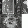
Fig. 6.1
Epidural angiolipoma in a 53-year-old woman. T2-weighted sagittal and axial images of the lumbar spine (a) show a 4 cm well-defined mass in the posterior epidural space at L1–2 level. Sagittal CT scan (b) shows internal fatty component. T1-weighted sagittal MR image (c) shows heterogeneous signal intensity of the lesion indicating mixed fatty and vascular components. Contrast-enhanced T1-weighted image (d) shows strong enhancement with some areas of fat signal suppression
6.2 Atypical Teratoid/Rhabdoid Tumor (ATRT)
- 1.
Epidemiology
Young children and infant, 7 months~17 years (more common in less than 3 years)
- 2.
Location
Most commonly in the brain, very rare in the spine
Intradural extramedullary, intramedullary
Cervical, thoracic, lumbar spine
- 3.
Characteristic imaging findings
Large heterogeneous mass
Internal hemorrhage
Diffuse contrast enhancement
CSF seeding
- 4.
Spectrum of imaging findings
- 5.
Differential diagnosis
Myxopapillary ependymoma and malignant peripheral nerve sheath tumor in extramedullary location
PNET, ependymoma, metastasis in intramedullary location
6.2.1 Illustrations: Atypical Teratoid/Rhabdoid Tumor (ATRT)

Fig. 6.2
Atypical teratoid/rhabdoid tumor (ATRT) in a 2-year-old child. Sagittal T2- and T1-weighted images (a–c) show a large heterogeneous mass at C1-C2-C3 levels with strong contrast enhancement. There is a focal cystic portion with suspected area of hemorrhage in the anteroinferior aspect of the tumor (white arrows). T2-weighted axial MR image (d) shows intradural extramedullary location of the tumor with cord compression and extradural extension into the left C1/C2 neural foramen
6.3 Chondroblastoma
- 1.
Epidemiology
20 years (9–62 years)
M > F
- 2.
Location
Very rare in spine, anywhere in the spine (most common in the thoracic spine)
Vertebral body and posterior elements
- 3.
Characteristic imaging findings
Expansive
Aggressive morphologic features with bony destruction and soft tissue mass
Calcification within the mass
- 4.
Spectrum of imaging findings
Secondary aneurysmal bone cyst
Bone marrow edema
- 5.
Differential diagnosis
Chondrosarcoma
Indistinguishable
Older ages (45–55 years)
6.3.1 Illustrations: Chondroblastoma

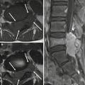
Fig. 6.3
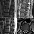
Chondroblastoma of the L3 vertebrae in a 25-year-old woman. T2-weighted sagittal and axial MR images (a, b) show pathologic compression fracture of the L3 vertebral body with multiple hemorrhagic fluid-fluid levels within the mass. Axial CT scan (c) shows internal fuzzy calcifications with cortical destruction at the right anterior aspect and paravertebral extension. The mass shows enhancing solid portions with adjacent bone marrow edema (d)
6.4 Epidural Hemangioma
- 1.
Epidemiology
Any age (mean age, 38 years; age range, 2–62 years)
M = F
- 2.
Location
Cavernous hemangioma: dorsal epidural space
Venous hemangioma: ventral epidural space, lumbar spine
- 3.
Characteristic imaging findings
Cavernous hemangioma: lobular contour, solid intense enhancement
- 4.
Spectrum of imaging findings
Manifested as epidural hematoma
Venous hemangioma: small cystic mass, T1-hyperintensity, ventral location
- 5.
Differential diagnosis
HIVD
T1-hypointensity, T2-hypointensity
Peripheral enhancement
Hematoma
No solid enhancement in acute stage
Schwannoma
Peripheral enhancement, less intense enhancement
Solid component
6.4.1 Illustrations: Epidural Hemangioma

Fig. 6.4
Epidural hemangioma in a 28-year-old woman. T2-weighted sagittal and axial MR images (a, b) show an extradural mass of high signal intensity at the left side of the T1 vertebra. T1-weighted MR image (c) shows intralesional high signal intensity suggesting internal hemorrhagic areas. T1-weighted axial MR image (d) shows mild enhancement

Fig. 6.5
Epidural hemangioma in a 30-year-old man. Lumbar spine MR images demonstrate a T2 hyperintense lesion with rim enhancement in the right anterior epidural space at the level of the upper body of L4 (a, b)

Fig. 6.6
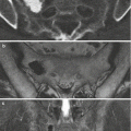
Spontaneous epidural hematoma without any tumor in a 36-year-old woman. Cervical spine MR images show an anterior epidural mass-like lesion extending from the levels of C7 to T4 of iso signal intensity on T1-weighted (a) and intermediate to high signal intensity on T2-weighted images (b). Nodular enhancement is seen centrally in the anterior spinal epidural lesion (c, d) indicating extravasation of contrast material by leaking vessels. Hyperacute to acute hematoma was diagnosed
6.5 Epithelioid Angiosarcoma
- 1.
Epidemiology
An extremely rare subtype of angiosarcoma, which is characterized by large cells with an epithelioid morphology
Middle aged and older
M = F
- 2.
Location
Rare in the spine
- 3.
Characteristic imaging findings
Feature of aggressive, high-grade bone tumor: destructive osteolytic mass with paravertebral extension, central necrosis, peripheral enhancement due to hyperperfusion
- 4.
Spectrum of imaging findings
Multicentricity (20–50%): multiple masses in a single bone
- 5.
Differential diagnosis
Epithelioid hemangioendothelioma
Difficult to differentiate
Less aggressive
Metastasis
Difficult to differentiate
Less hypervascular
6.5.1 Illustrations: Epithelioid Angiosarcoma


Fig. 6.7
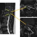
Epithelioid angiosarcoma in a 73-year-old man. T2 SPAIR and T1-weighted sagittal MR images (a, b) show a heterogeneous signal mass in the L1 vertebral body with extensive bone marrow edema. T2-weighted axial MR images (c, d) show internal tortuous low signal flow voids (white arrows). Pre-surgical angiography for tumor embolization (e) shows marked hypervascularity within and around the tumor
6.6 Epithelioid Hemangioendothelioma
- 1.
Epidemiology
Vascular tumor, intermediate between hemangiomas and conventional angiosarcomas
Very rare in the spine
Middle aged and older
M = F
- 2.
Location
Cervical, lumbar
- 3.
Characteristic imaging findings
Nonspecific vascular tumor
Osteolytic mass, hypervascularity, single or multiple
- 4.
Spectrum of imaging findings
Distant metastasis (20–30%)
- 5.
Differential diagnosis
Hemangioma
Less aggressive
Can be cystic
Epidural hematoma
Angiosarcoma
More aggressive
More bony destruction and paravertebral extension
Metastasis
Less hypervascular
6.6.1 Illustrations: Epithelioid Hemangioendothelioma

Fig. 6.8
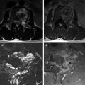
Epithelioid hemangioendothelioma in a 55-year-old man. T2-weighted sagittal MR image (a) shows a 2 cm intramedullary mass of high signal intensity at the level of T10 vertebra. Extensive spinal cord edema is combined. Contrast-enhanced T1-weighted axial MR image (b) shows an exophytic intramedullary mass in the left side with intense homogenous enhancement. Contrast-enhanced T1-weighted sagittal MR image (c) shows a vascular pedicle in the inferior aspect of the tumor (white arrow). Hypervascular tumor staining from the left T10 spinal artery (black arrow) is demonstrated on angiography (d, e)
6.7 Ganglioglioma
- 1.
Epidemiology
Children, young adult
M = F
- 2.
Location
Intramedullary location
Cervical spinal cord > thoracic spinal cord
Related posts:

Stay updated, free articles. Join our Telegram channel

Full access? Get Clinical Tree