Chapter 16 Recognizing Abnormal Calcifications and Their Causes
 Soft tissue calcifications lend themselves to an intuitive approach that ties together a diverse group of diseases. While this chapter focuses primarily on abdominal calcifications, the same principles and approach apply to dystrophic calcifications found anywhere in the body.
 Dystrophic calcification is soft tissue calcification that occurs in tissue that is already abnormal. (Metastatic calcification, by contrast, is calcification in otherwise normal soft tissues due to disturbances in calcium/phosphorus metabolism.)
 On conventional radiographs, the nature of most calcifications can be determined by examining their pattern of calcification and their anatomic location.
 The same principles hold true for CT evaluation of calcifications except that the anatomic location is usually easier to determine on CT.
Rimlike Calcification
 Examples of structures that manifest rimlike calcifications:
• Aneurysms
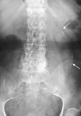
• Aortic aneurysm. CT and US are used to identify and characterize abdominal aortic aneurysms. On conventional radiographs, such aneurysms are most easily recognized on a lateral radiograph of the lumbar spine. The abdominal aorta should normally measure <3 cm in diameter (Fig. 16-2).
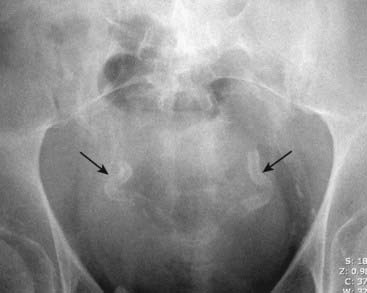
• Saccular organs, such as the gallbladder or urinary bladder
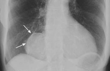
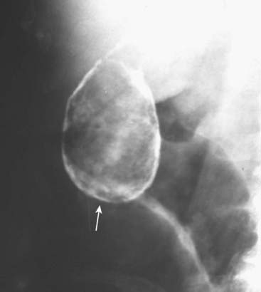
• Porcelain gallbladder—an uncommon entity (named after the gross appearance of the gallbladder, which resembles porcelain) that occurs with chronic inflammation of and stasis in the gallbladder and is associated with gallstones and an increased incidence of carcinoma of the gallbladder (Fig. 16-3).
TABLE 16-1 RIMLIKE CALCIFICATIONS
| Organ of Origin | Remarks |
|---|---|
| Renal cyst | Thick and irregular calcifications, though uncommon, may indicate the presence of renal cell carcinoma. |
| Splenic cysts | May be a manifestation of hydatid cyst, old trauma, or prior infection. |
| Aortic aneurysms | Occurs more often in diabetics with advanced atherosclerosis. |
| Gallbladder | Associated with chronic stasis; called porcelain gallbladder for its gross appearance; higher incidence of carcinoma of the gallbladder. |


