Chapter 15 Recognizing Extraluminal Air in the Abdomen
 Recognition of extraluminal air is an important finding that can have an immediate effect on the course of treatment.
 Air is normally not present in the peritoneal or extraperitoneal spaces, bowel wall, or biliary system.
Signs Of Free Intraperitoneal Air
Air Beneath the Diaphragm
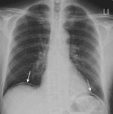
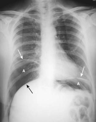
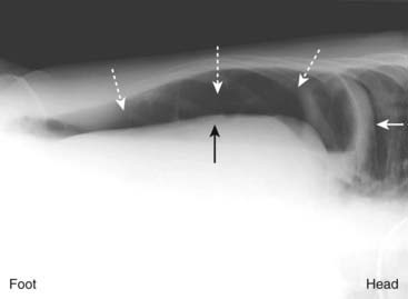
 Air will rise to the highest part of the abdomen. Therefore, in the upright position, free air will usually reveal itself under the diaphragm as a crescentic lucency that parallels the undersurface of the diaphragm (Fig. 15-1).
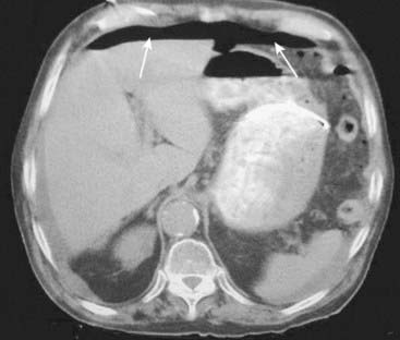
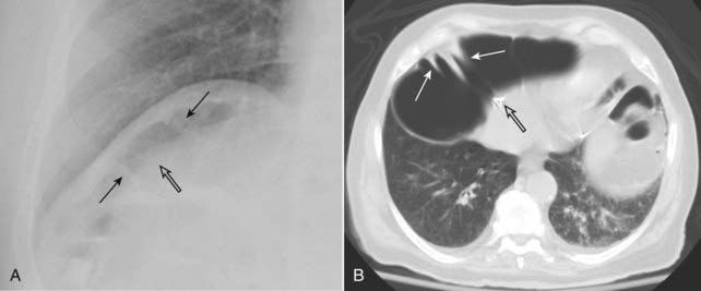
 While free air is best demonstrated on CT scans of the abdomen because of its greater sensitivity in detecting very small amounts of free air (Fig. 15-3), most surveys of the abdomen begin with conventional radiographs. Conventional radiographs serve as an important screening tool on which many previously unsuspected cases of free air are discovered.
![]() On conventional radiographs, free air is best demonstrated with the x-ray beam directed parallel to the floor (i.e., a horizontal beam) (see Figs. 13-14 and 13-15). Small amounts of free air will not be visible on supine radiographs.
On conventional radiographs, free air is best demonstrated with the x-ray beam directed parallel to the floor (i.e., a horizontal beam) (see Figs. 13-14 and 13-15). Small amounts of free air will not be visible on supine radiographs.
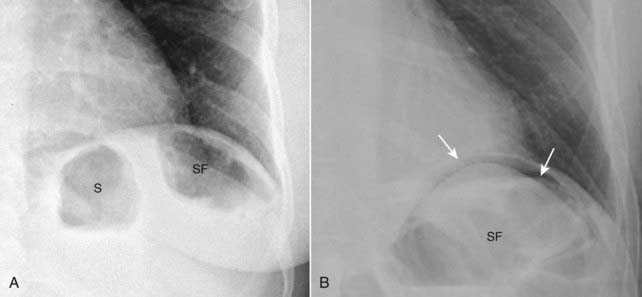
 Free air is easier to recognize under the right hemidiaphragm because only the soft tissue density of the liver is usually located there. Free air is more difficult to recognize under the left hemidiaphragm because air-containing structures, such as the fundus of the stomach and the splenic flexure, already reside in that location and may mask the presence of free air (Fig. 15-4).
 If the patient is unable to stand or sit upright, then a view of the abdomen with the patient lying on his or her left side taken with a horizontal x-ray beam may show free air rising above the right edge of the liver. This is the left lateral decubitus view of the abdomen (Fig. 15-5).
• Occasionally, colon may be interposed between the dome of the liver and the right hemidiaphragm and may be mistaken for free air, unless a careful search is made for the presence of haustral folds characteristic of the colon (Fig. 15-6).
Visualization of Both Sides of the Bowel Wall
 In the normal abdomen, we visualize only the air inside the lumen of the bowel, not the wall of the bowel itself. This is because the wall is soft tissue density and surrounded by tissue of the same density.
 Introduction of air into the peritoneal cavity enables us to visualize the wall of the bowel itself since the wall is now surrounded on both inside and outside by air.
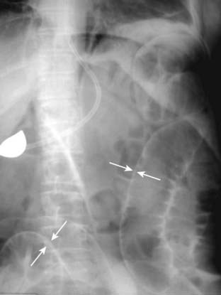
• The ability to see both sides of the bowel wall is a sign of free intraperitoneal air called Rigler sign. Rigler sign usually requires large amounts of free air in order to be present (Fig. 15-7).
• The sign can be seen on supine, upright, or prone films of the abdomen so long as an adequate amount of free air is present.
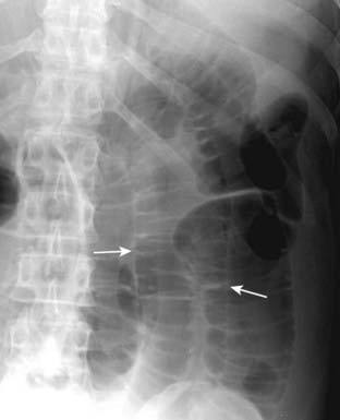
![]() Pitfall: When dilated loops of small bowel overlap each other, they may occasionally produce the mistaken impression that you are seeing both sides of the bowel wall (Fig. 15-8).
Pitfall: When dilated loops of small bowel overlap each other, they may occasionally produce the mistaken impression that you are seeing both sides of the bowel wall (Fig. 15-8).
Visualization of the Falciform Ligament
 The falciform ligament courses over the free edge of the liver anteriorly just to the right of the upper lumbar spine. It contains a remnant of the obliterated umbilical artery.
Stay updated, free articles. Join our Telegram channel

Full access? Get Clinical Tree


