Chapter 2 Recognizing Normal Chest Anatomy and a Technically Adequate Chest Radiograph
 In order to become more comfortable interpreting chest radiographs, you first need to be able to recognize fundamental, normal anatomy so you can differentiate it from what is abnormal.


The Normal Frontal Chest Radiograph

 Vessels and bronchi: normal lung markings
 Pleura: normal anatomy
The Lateral Chest Radiograph
 As part of the standard two-view chest examination, patients usually have an upright, frontal chest radiograph and an upright, left lateral view of the chest.

 Why look at the lateral chest?
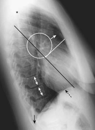
Figure 2-2 Normal left lateral chest radiograph.
A clear space is present behind the sternum (solid white arrow). The hila produce no discrete shadow (white circle). The vertebral bodies are approximately of equal height and their endplates are parallel to each other (double white arrows). The posterior costophrenic angles (solid black arrow) are sharp. Notice how the thoracic spine appears to become blacker (darker) from the shoulder girdle (black star) to the diaphragm because there is less dense tissue for the x-ray beam to traverse at the level of the diaphragm. The heart normally touches the anterior aspect of the left hemidiaphragm and usually obscures (silhouettes) it. The superior surface of the right hemidiaphragm is frequently seen continuously from back to front (dotted black arrow) because it is not obscured by the heart. Notice the normal space posterior to the heart and anterior to the spine; this will be important in assessing cardiomegaly (Chapter 9). The black line represents the approximate location of the major or oblique fissure; the white line is the approximate location of the minor or horizontal fissure. Both are visible because they are seen en face on the lateral view.
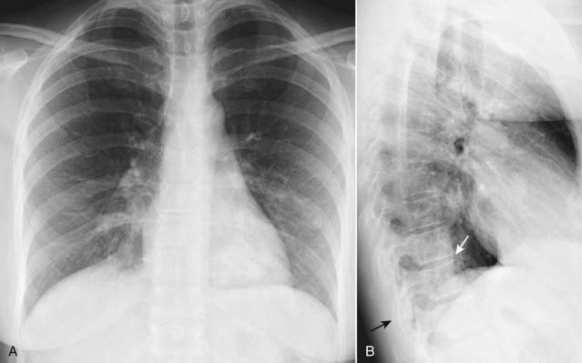
Figure 2-3 The spine sign.
Frontal (A) and lateral (B) views of the chest demonstrate airspace disease on the lateral film (B) in the left lower lobe that may not be immediately apparent on the frontal film (look closely at A and you may see the pneumonia in the left lower lobe behind the heart). Normally, the thoracic spine appears to get “blacker” as you view it from the neck to the diaphragm because there is less dense tissue for the x-ray beam to traverse just above the diaphragm than in the region of the shoulder girdle (see also Fig. 2-2). In this case, a left lower lobe pneumonia superimposed on the lower spine in the lateral view (solid white arrow) makes the spine appear “whiter” (more dense) just above the diaphragm. This is called the spine sign. Note that on a well-positioned lateral projection, the right and left posterior ribs almost superimpose on each other (solid black arrow), a sign of a true lateral.
Five Key Areas on the Lateral Chest X-Ray (Fig. 2-2 and Table 2-1)


TABLE 2-1 THE LATERAL CHEST: A QUICK GUIDE OF WHAT TO LOOK FOR
| Region | What You Should See |
|---|---|
| Retrosternal clear space | Lucent crescent between sternum and ascending aorta |
| Hilar region | No discrete mass present |
| Fissures | Major and minor fissures should be pencil-point thin, if visible at all |
| Thoracic spine | Rectangular vertebral bodies with parallel end plates; disk spaces maintain height from top to bottom of thoracic spine |
| Diaphragm and posterior costophrenic sulci | Right hemidiaphragm slightly higher than left; sharp posterior costophrenic sulci |
The Retrosternal Clear Space
 Normally, a relatively lucent crescent is present just behind the sternum and anterior to the shadow of the ascending aorta.
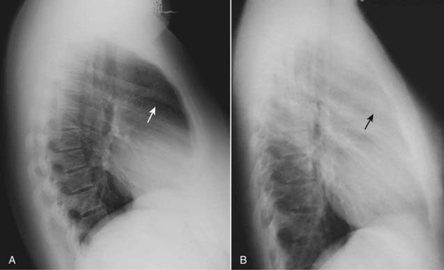
 Pitfall: Be careful not to mistake the soft tissue of the patient’s superimposed arms for “filling-in” of the clear space. Although patients are asked to hold their arms over their head for a lateral chest exposure, many are too weak to raise their arms.
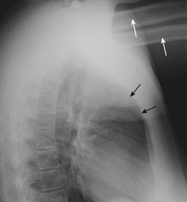
Figure 2-5 Arms obscure retrosternal clear space.
In this example, the patient was not able to hold her arms over her head for the lateral chest examination, as patients are instructed to do in order to eliminate the shadows of the arms from overlapping the lateral chest. The humeri are clearly visible (solid white arrows) so even though the soft tissue of the patient’s arms appears to fill in the retrosternal clear space (solid black arrows), this should not be mistaken for an abnormality such as anterior mediastinal adenopathy (see Fig. 2-4).
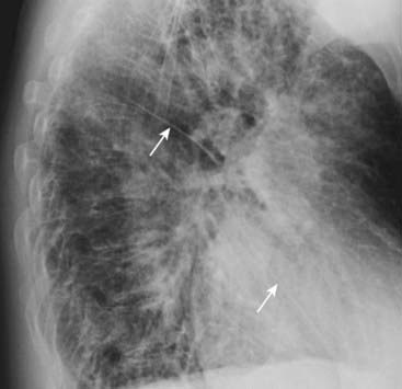
The Hilar Region
 The hila may be difficult to assess on the frontal view, especially if both hila are slightly enlarged, since comparison with the opposite normal side is impossible.
 When there is a hilar mass, such as might occur with enlargement of hilar lymph nodes, the hilum (or hila) will cast a distinct, lobulated masslike shadow on the lateral radiograph (Fig. 2-6).
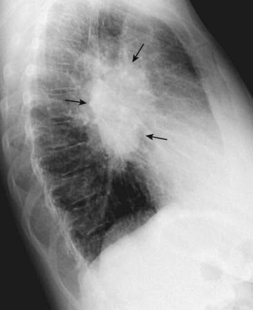
The Fissures
 On the lateral film, both the major (oblique) and minor (horizontal) fissures may be visible as a fine, white line (about as thick as a line made with the point of a sharpened pencil).
 When a fissure contains fluid or develops fibrosis from a chronic process, it will become thickened (Fig. 2-7).