Chapter 4 Recognizing the Causes of an Opacified Hemithorax
 Mr. Smith, age 73, presents to the Emergency Department very short of breath. His frontal chest radiograph is shown in Figure 4-1.
 As you can see, Mr. Smith’s right hemithorax is almost completely opaque.
• Mr. Smith’s treatment will vary greatly depending on whether he has atelectasis (which may require emergent bronchoscopy), a large effusion (which may require emergent thoracentesis), or pneumonia (which would require starting antibiotics).

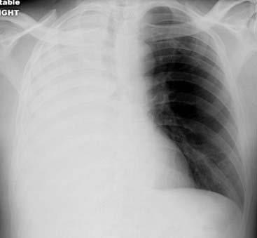
Figure 4-1 Mr. Smith comes into the Emergency Department short of breath. This is his frontal chest radiograph.
Atelectasis of the Entire Lung
 Atelectasis of an entire lung usually results from complete obstruction of the right or left main bronchus.
• With bronchial obstruction, no air can enter the lung. The remaining air in the lung is absorbed into the bloodstream through the pulmonary capillary system.

 In obstructive atelectasis, even though there is volume loss within the affected lung, the visceral and parietal pleura almost never separate from each other.
• That is an important fact about atelectasis and is sometimes confusing to beginners who try to picture atelectasis and a pneumothorax as both producing collapse of a lung without understanding why they look completely different (Fig. 4-2 and Table 4-1).
• Because the visceral and parietal pleura do not separate from each other in atelectasis, mobile structures in the thorax are “pulled” toward the side of the atelectasis producing a shift (movement) of certain mobile thoracic structures toward the side of opacification.
• The most visible mobile structures in the thorax are the heart, the trachea, and the hemidiaphragms.
• In obstructive atelectasis, one or all of these structures will shift toward the side of opacification (toward side of volume loss) (Fig. 4-3).
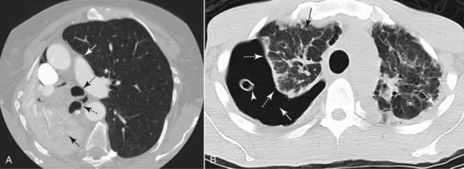
TABLE 4-1 PNEUMOTHORAX VERSUS OBSTRUCTIVE ATELECTASIS
| Feature | Pneumothorax | Obstructive Atelectasis |
|---|---|---|
| Pleural space | Air in the pleural space separates the visceral from the parietal pleura | The visceral and parietal pleura do not separate from each other |
| Density | The pneumothorax itself will appear “black” (air density); the hemithorax may appear more lucent than normal | Atelectasis is the absence of air in the lung; the hemithorax will appear more opaque (“whiter”) than normal |
| Shift | The heart or trachea never shift toward the side of a pneumothorax | The heart and trachea almost always shift toward the side of the atelectasis |
Related posts:
Stay updated, free articles. Join our Telegram channel

Full access? Get Clinical Tree


