Chapter 17 Recognizing the Imaging Findings of Trauma
 Trauma is the leading cause of death, hospitalization, and disability in Americans from the age of 1 year through age 45. The major imaging findings of most organ system’s trauma will be discussed as a group in this chapter. Table 17-1 summarizes some of the traumatic injuries that are discussed in other chapters.
TABLE 17-1 OTHER MANIFESTATIONS OF TRAUMA
| Injury | Discussed in |
|---|---|
| Pleural effusion/hemothorax | Chapter 6 |
| Aspiration | Chapter 7 |
| Pneumothorax, pneumomediastinum and pneumopericardium | Chapter 8 |
| Fractures and dislocations | Chapter 22 |
| Head trauma | Chapter 25 |
Chest Trauma
 Chest injuries in trauma patients are very common and are responsible for one out of four trauma-related deaths. The overwhelming majority of chest trauma is the result of motor vehicle accidents.
Rib Fractures
 The severity of underlying visceral injury is usually more important than the rib fractures themselves, but their presence might provide clues to unsuspected pathology.
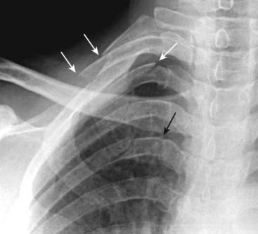
 Fractures of the first three ribs are relatively uncommon and, if they occur following blunt trauma, indicate a sufficient amount of force to produce other internal injuries (Fig. 17-1).


Pulmonary Contusions
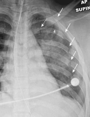
 Pulmonary contusions are the most frequent complications of blunt chest trauma. They represent hemorrhage into the lung, usually at the point of impact.
 Recognizing a pulmonary contusion
• The history of trauma is of paramount importance as contusions present as airspace disease that is indistinguishable from other airspace diseases like pneumonia or aspiration.


Pulmonary Lacerations (Hematoma or Traumatic Pneumatocele)
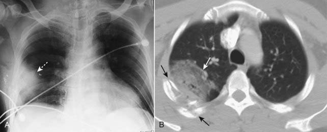
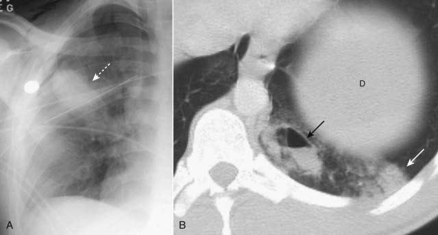
 Pulmonary hematomas result from a laceration of the lung parenchyma and, as such, may accompany more severe blunt trauma or penetrating chest trauma.

 Recognizing a pulmonary laceration
• Their appearance will depend on whether they contain blood and, if so, how much blood fills the laceration.
• If they are partially filled with blood and partially filled with air, they may contain a visible air-fluid level or demonstrate a crescent sign as the blood begins to form a clot and pull away from the wall of the laceration.

Aortic Trauma
 Trauma to the aorta is most frequently the result of deceleration injuries in motor vehicle accidents. Although the survival rates are improving, most patients with rupture of the thoracic aorta die before reaching the hospital and, of those who survive an aortic injury, the likelihood of death increases the longer the abnormality remains untreated. Only those with incomplete tears in which the adventitial lining prevents exsanguination (producing a pseudoaneurysm) survive to be imaged.

 Recognizing aortic trauma
• Findings seen on conventional radiographs of the chest are the same as those discussed under Aortic Dissection in Chapter 9. A completely normal chest radiograph has a relatively high negative predictive value for aortic injury, but an abnormal chest x-ray has a relatively low positive predictive value


Related posts:
Stay updated, free articles. Join our Telegram channel
Full access? Get Clinical Tree
