Rectus Sheath Hematoma
Ho Chia Ming
CLINICAL HISTORY
73-year-old man with a history of heterozygous factor V Leiden deficiency and a recent history of deep vein thrombosis treated with anticoagulation, presented with an enlarging abdominal mass.
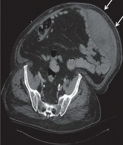 FIGURE 19A |
FINDINGS
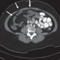
Figure 19A: Axial contrast-enhanced CT through the pelvis shows marked thickening of the left rectus abdominis muscle with a biconvex lentiform shape and heterogeneous contents that are slightly hyperattenuated, compatible with rectus sheath hematoma (arrows).
DIFFERENTIAL DIAGNOSIS
Solitary fibrous tumor, desmoid, neurofibroma, metastases, lymphoma, sarcoma, abdominal wall abscess, rectus sheath hematoma.
DIAGNOSIS
Rectus sheath hematoma.
DISCUSSION
A hematoma in the sheath of the rectus abdominis muscle results from injury to the superior or inferior epigastric arteries or rectus muscle. Causes of rectus sheath hematoma include trauma, abdominal surgery, and vigorous abdominal muscle contractions. Spontaneous nontraumatic hematoma occurs most commonly because of anticoagulation. Signs of rectus sheath hematoma include ecchymosis in the periumbilical region or the flanks, and a tender abdominal mass on examination, which remains or becomes more conspicuous (Fothergill sign) or tender (Camett sign) on tensing the abdominal wall.
Related posts:


Stay updated, free articles. Join our Telegram channel

Full access? Get Clinical Tree


