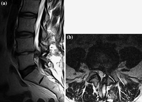
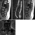
Fig. 1
a–b. FSE T2 sagittal (a), and axial (b). L4–L5 recurrent left paramedian-intraforaminal hernia that migrates caudally, characterized by herniated nucleus pulposus (hydrated and T2 hyperintense); absence of radicular conflict
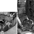
Preoperative Imaging After 10 Months
 < div class='tao-gold-member'>


Only gold members can continue reading. Log In or Register to continue
Related posts:

Stay updated, free articles. Join our Telegram channel
Full access? Get Clinical Tree
