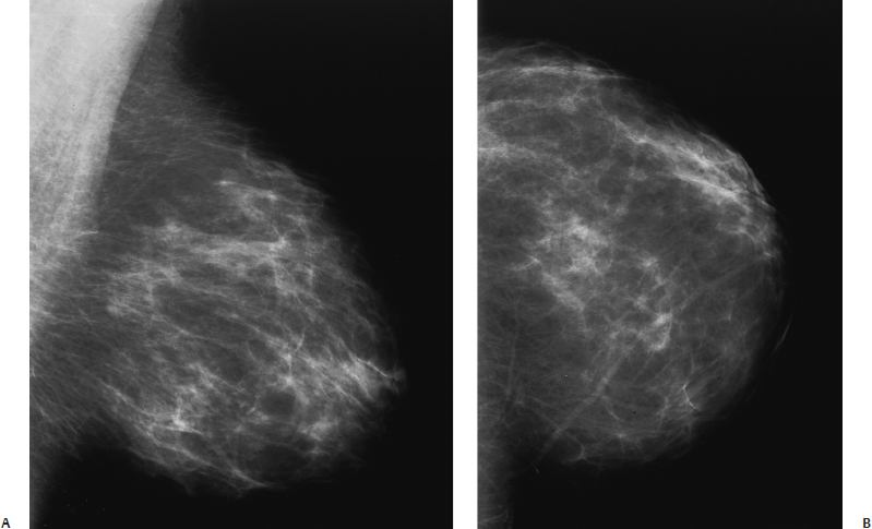
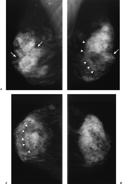
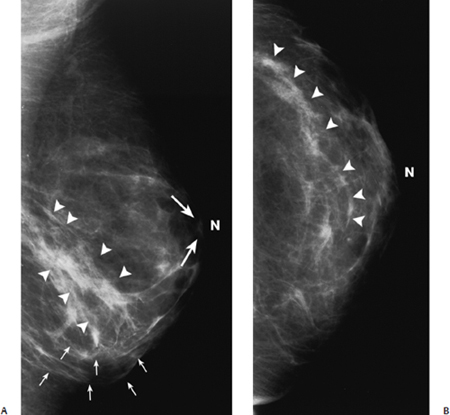
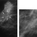
27 Reduction Mammoplasty A 61-year-old woman had reduction mammoplasty 2 years ago. • Bilateral breasts: bilateral reduction scars, otherwise normal Fig. 27.1 Normal mammogram prior to reduction mammoplasty. (A) Left MLO mammogram. (B) Left CC mammogram. Fig. 27.2 Normal mammogram 2 years after reduction mammoplasty. The nipple (N) is higher in position, and the contour of the breast is flatter. Architectural distortion has resulted in swirled lines (small arrows); disruption of the normal subareolar ductal lines (large arrows); and increase in the density of the inferior breast, which in this patient is associated with straight parenchymal bands (arrowheads). (A) Left MLO mammogram. (B) Left CC mammogram. • Changes from reduction mammoplasty • BI-RADS assessment category 2, benign finding • The various reduction mammoplasty procedures involve removing tissue from the inferior breast. The residual upper breast tissue is brought together in the midline to reform a smaller breast, and the nipple areolar complex is transposed superiorly. After these procedures, mammograms exhibit the following findings: (1) elevation of the nipple with flattening of the breast contour, (2) architectural distortion, (3) fat necrosis, (4) dystrophic calcifications, and (5) skin thickening. Jackson VP. Reduction mammoplasty. In: Bassett LW, Jackson VP, Jahan R, Fu YS, Gold RH, eds. Diagnosis of Diseases of the Breast. Philadelphia: WB Saunders; 1997:581–587 An 85-year-old woman presents for screening mammogram. • Bilateral breasts: reduction mammoplasty scars, otherwise normal Fig. 27.3 Bilateral normal mammograms after reduction. Architectural distortion from reduction has resulted in asymmetry in both the anterior and posterior contours of the breasts. There are unusual indentations in the parenchymal outline (arrows
Case 27.1: Architectural Distortion
Case History
Physical Examination
Mammogram (Figs. 27.1 and 27.2)
Pathology
Management
Pearls and Pitfalls
Suggested Reading
Case 27.2: Architectural Distortion
Case History
Physical Examination
Mammogram (Fig. 27.3)
![]()
Stay updated, free articles. Join our Telegram channel


Full access? Get Clinical Tree


