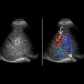
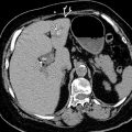
KEY FACTS
Terminology
- •
Global or focal renal hypoperfusion → tissue ischemia and eventually, parenchymal loss
Imaging
- •
Sonographic diagnosis is difficult; evaluation by CT/MR more common
- •
± alteration in grayscale appearance with ↓ corticomedullary differentiation
- •
Focally diminished or absent Doppler flow
- •
May involve all or part of 1 kidney
- •
Insults to accessory renal arteries tend to cause polar infarcts
- •
Wedge-shaped, corresponding to vascular territory in kidney
- •
May be hypoechoic or hyperechoic, depending on timing
- •
When focal, tends to be wedge-shaped and extend all the way from hilum to capsule
- •
Focal or global loss of parenchymal flow
Pathology
- •
Arterial disease: Trauma, atherosclerosis, vasculitis, dissection
- •
Embolism: Endocarditis, arrhythmias with clot
- •
Thrombosis: Trauma or hypercoagulability
- •
Iatrogenic: Small polar arteries may be sacrificed in AAA repair or transplant harvest
Diagnostic Checklist
- •
Clinical context will usually help to exclude the other items in differential diagnosis
- •
Look for ancillary signs of infection or trauma on CT exams to narrow the differential
- •
Small polar infarcts are common after endovascular repair of AAA
Scanning Tips
- •
Scanning in lateral decubitus or oblique position in coronal scan plane through posterior axillary line results in shorter Doppler distance and improved color Doppler sensitivity
- •
Optimize settings to detect slow flow in color and power Doppler
- •
Make sure to scan kidneys in horizontal presentation such that both upper pole and lower poles are located at similar depth from transducer; this will ensure entire kidney is interrogated equally with color Doppler
- •
Reduce size of Doppler box while still including kidney to optimize visualization of vascular defects


