KEY FACTS
Imaging
- •
Usually small and round, may be wedge-shaped
- •
Usually cortical; rarely disrupting renal contour or capsule
- ○
Large, exophytic metastases may be encountered
- ○
- •
Hypoechoic, hyperechoic, or sonographically occult
- •
Occasionally infiltrative
- •
Perinephric infiltration from tumor extension or hemorrhage may be seen (melanoma)
- •
Mostly avascular or hypovascular using low-flow Doppler settings
- •
Melanoma (and other hypervascular primary tumors) metastasis can be hypervascular, simulating renal carcinoma
Top Differential Diagnoses
- •
Primary renal malignancy
- •
Renal angiomyolipoma
- •
Renal cysts (complex)
- •
Renal lymphoma or leukemia
- •
Renal infection
- •
Renal infarction
Clinical Issues
- •
May have hematuria or microhematuria (12-31%)
- •
Most are clinically occult and found on imaging or at autopsy
- •
Most common malignant renal tumor at autopsy (7-13% of autopsies); 20% of patients dying of disseminated malignancy
- •
Most patients have metastatic tumor at other locations
- •
Lung cancer most common primary site followed by breast, gastric cancer, melanoma, and lymphoma
- •
Prognosis very poor
Scanning Tips
- •
Metastases may be subtle on ultrasound; use color Doppler at low-flow settings
- •
Presence of disseminated malignancy suggests metastases as cause of abnormal renal ultrasound
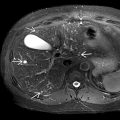
 representing metastatic melanoma. This cannot be distinguished from renal cell carcinoma.
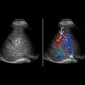
 . While the grayscale features are indistinguishable from renal cell carcinoma, metastases are typically hypovascular compared to renal cell carcinoma.