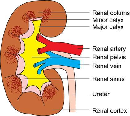
Proliferation of fat within the kidney represents a spectrum of disorders ranging from mild fatty deposition within the renal sinus with preserved parenchyma (renal sinus lipomatosis [RSL]) to a severe variety with fatty proliferation involving not only the renal sinus but also extending to the hilum and perinephric region with underlying parenchymal atrophy (renal replacement lipomatosis [RRL]). RSL refers to a condition where there is excessive nontumorous fatty tissue within the renal sinus. Along the medial border of the kidney, the extension of perinephric space forms the deep recess – the renal sinus (Fig. 10.12.2.6.1). Laterally the renal sinus is surrounded by the kidney parenchyma. The fatty tissue within the renal sinus is continuous with the perirenal fat through crevices between the hilar structures. Within the renal sinus are located the major branches of the renal artery and vein along with the major and minor calices of the collecting system. The remainder of the sinus is filled with lymphatic channels, adipose tissue, nerve fibres of the autonomic nervous system and varying quantities of fibrous tissue. A nontumorous increase in the sinus fat is termed as RSL.
6. Renal sinus lipomatosis
Anatomy
Aetiology
Related posts:


Stay updated, free articles. Join our Telegram channel

Full access? Get Clinical Tree


