KEY FACTS
Terminology
- •
Clot formation in renal vein
Imaging
- •
Unilateral > bilateral
- •
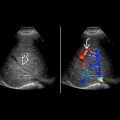
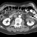
Kidney enlarged acutely in 75% cases
- •
Renal vein dilated acutely
- •
Possible inferior vena cava (IVC) thrombus extension
- •
Altered renalarteryspectral waveforms
- ○
↑ systolic pulsatility (narrow, sharp systolic peaks)
- ○
Persistent retrograde diastolic flow
- ○
Pathology
- •
Nephrotic syndrome: Most common cause of renal vein thrombosis in adults
- •
Hypovolemia/renal hypoperfusion: Most common cause of renal vein thrombosis in children
- •
Risk in neonates is associated with fetal distress, perinatal asphyxia, diabetic mothers, and volume contraction
Clinical Issues
- •
Outcome depends on cause, time to diagnosis, duration of occlusion, recanalization, collateralization
- •
Prognosis overall good; frequent spontaneous recovery
- •
Anticoagulation: Heparin then Coumadin or low-molecular-weight heparin for maintenance
- •
Thrombolysis/surgical thrombectomy: Heroic measure for life-threatening situations
- •
Suprarenal caval filter (IVC thrombus)
Scanning Tips
- •
Renal vein can be scanned from flank or epigastric window; flank window is more frequently used because it is less affected by body habitus and has fewer surrounding vessels
- •
Do not mistake splenic vein for left renal vein (when using epigastric window)
- ○
Splenic vein anterior to superior mesenteric artery
- ○
Left renal vein posterior to superior mesenteric artery
- ○
 .
.


