Retropharyngeal Abscess
Benjamin Y. Huang
CLINICAL HISTORY
5-month-old infant with a 2-week history of intermittent fevers, now presenting with left neck and chin swelling.
FINDINGS
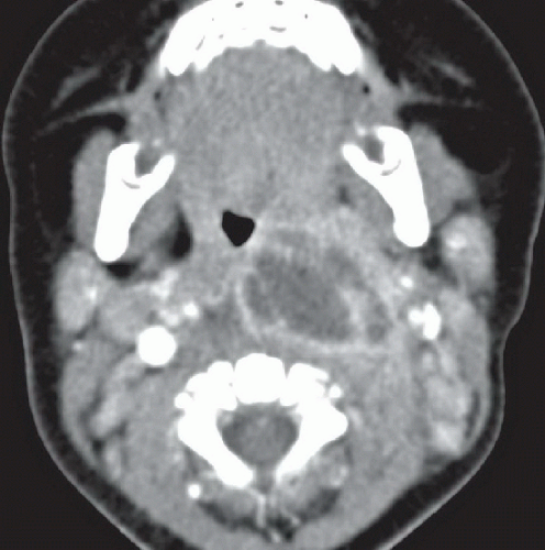
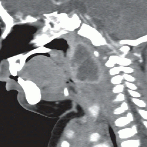
Figures 57A and 57B: Axial (Fig. 57A) and sagittal (Fig. 57B) contrast-enhanced CT images reveal a large, ovoid fluid collection in the left lateral retropharyngeal space that demonstrates an irregular enhancing rim and internal septations. The fluid collection significantly displaces the oropharyngeal airway anteriorly and to the right. There is also enhancing cervical lymphadenopathy, which is slightly more pronounced on the left.
DIFFERENTIAL DIAGNOSIS
The primary diagnostic consideration given the history and imaging findings is a retropharyngeal abscess. Tonsillar and peritonsillar abscesses are generally centered in the tonsils along the lateral pharyngeal wall and may extend laterally into the parapharyngeal space. Suppurative retropharyngeal lymphadenitis can also appear as a peripherally enhancing fluid collection in the lateral retropharyngeal space; however, these are typically smaller and less centrally located than abscesses. Sterile retropharyngeal effusions/edema also occur in the setting of upper respiratory tract infections and also appear as fluid collections in the retropharyngeal space; however, these effusions do not show peripheral enhancement, are typically midline, and demonstrate a more lentiform appearance on the sagittal images. Lymphatic and mixed vascular malformations can also appear as low-density masses and involve the retropharyngeal space; however, the pattern of enhancement in mixed vascular malformations is typically not isolated to the periphery of the lesions, and the clinical history in this case does not support the diagnosis.
Stay updated, free articles. Join our Telegram channel

Full access? Get Clinical Tree


