(1)
Department of Clinical Radiology, Amiri Hospital – Kuwait City, Kuwait City, Kuwait
6.3 Gout Arthritis
6.4 CPPD and HADD
6.5 Osteoarthritis
6.10 Behçet Disease
6.10.1 Clinical Criteria to Diagnose BD Require a Combination of Three or More of the Following Findings
6.1 Rheumatoid Arthritis
Rheumatoid arthritis (RA) is a chronic, multisystemic, nonspecific inflammatory disease with unknown etiology that primarily affects the joints and the skeletal muscles.
RA is diagnosed by qualifying certain criteria. Four of the following criteria should be present for more than 6 months to fulfill the diagnosis of RA:
Morning stiffness that lasts at least an hour before maximal improvement
Soft-tissue swelling and arthritis in a bilateral symmetrical fashion of at least three joints (polyarthritis)
Swelling of the metacarpophalangeal, proximal phalangeal, or wrist joints
Subcutaneous rheumatoid nodules
A positive test for rheumatoid factor
Radiographic signs of RA
In joints, the main pathology of RA is related to synovial inflammation and proliferation. The normal synovium is attached to the inner joint capsule in synovial joints. The articulating surface of the joint is covered with cartilage except for a small region at the insertion of the joint capsule. This area is covered only by synovium, and it is called the “bare area.” Synovial inflammation and bone erosions start from this area in RA. The granulation tissue (pannus) that results from the chronic synovial inflammation adheres and extends to the articular cartilage and the subchondral surface. This extension causes bone resorption and articular adhesions that may ossify and leads to bony fusion. Intra-articular loose bodies may develop as a consequence of the inflammatory process. The loose intra-articular bodies are composed of destroyed cartilage or hypertrophied synovium.
RA affects mainly women between 30 and 40 years of age. Any joint in the body can be affected by RA, but the disease often affects the small joints of the hands excluding the terminal phalanges. Wrists, knees, and feet are also commonly affected by RA. Patients often present with morning stiffness, small joints swelling due to tenosynovitis, muscular pain, and stiffness commonly after a period of immobilization. Extra-articular manifestations of RA make the disease mixed in its early stage with systemic lupus erythematosus (SLE). SLE, however, may be seen in patients with chronic RA.
Nervous system manifestations in RA can be categorized into four groups: central nervous system rheumatoid nodules, cerebral vasculitis, cervical myelopathy due to atlantoaxial subluxation, and peripheral neuropathy. The atlantoaxial subluxation may lead to the development of double crush syndrome. The double crush hypothesis refers to the concept that a single lesion in the course of a nerve predisposes that nerve to a second lesion further along its course. This phenomenon is often observed in patients with thoracic outlet syndrome, where compression of the brachial plexus can result in the development of carpal tunnel syndrome. Median nerve neuritis (carpal tunnel syndrome), Raynaud’s phenomenon, and hyperemia of the palms (liver palms) may be seen in cases of rheumatoid peripheral neuropathy and sympathetic nervous system hyperactivity. Moreover, the cervical manifestations of RA can be seen as rheumatoid discitis and myelopathy due to thickening of the dura. Rheumatoid discitis arises due to annulus fibrosis and replacement of the normal intravertebral disk by rheumatoid pannus.
Rheumatoid nodules (10–20 %) are seen in juxta-articular surfaces or on the extensor surfaces of the arm and elbows, especially the olecranon surface. Rheumatoid nodules consist histologically of three zones: a central zone of necrotic tissue, a middle zone of histiocytes and monocytes, and an outer zone of chronic inflammatory granulation tissue. They can affect any part of the body including the heart, larynx, eye, Achilles’ tendon, and lungs. The presence of rheumatoid nodules is indicative of severe disease. An uncommon complication of rheumatoid nodule includes breakdown of the overlying skin and discharge of the content (fistulous rheumatism). Rarely, rheumatoid nodules may present as linear, elongated, cord-like subcutaneous bands.
Moderate hypochromic normocytic anemia can be found in chronic RA due to anemia of chronic disease. Diseases that can be associated with RA are psoriasis (8 %), ulcerative colitis, amyloidosis, and asthma. Lymphadenopathy can be seen in cases of RA.
Rheumatoid nodulosis (RN) is a rare, benign RA variant characterized by the presence of subcutaneous rheumatoid nodules with absence of synovitis, absence of systemic manifestations in benign course, and mild radiological findings. Classically, the presence of rheumatoid nodule is a sign of advanced RA with poor prognosis. As a rule, the benign course of the disease and the mild radiological findings are what differentiate RA from RN. RN is found in up to 25 % of classical RA cases and presents between ages 30 and 50 years. RN has a male predominance (81 %), whereas RA has a female predominance. The subcutaneous rheumatoid nodules in RN appear at the onset of clinical symptoms and are usually observed over bony surfaces like the back of the hands and the olecranon (Fig. 6.1.1).
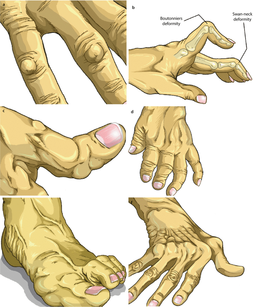
Fig. 6.1.1
An illustration demonstrates different manifestations of rheumatoid arthritis (RA) in the hand: (a) rheumatoid nodulosis, (b) Boutonnière and swan-neck deformities, (c) hitchhiker thumb deformity, (d) opera-glass hand, (e) hammertoe deformity, and (f) ulnar deviation
CNS manifestations of RA include vasculitis, pachymeningitis, leptomeningitis, rheumatoid nodules formation, stroke, seizures, and encephalopathy. The diagnosis of cerebral manifestation of RA must be supported by high titers of RF and anticyclic citrullinated peptide (anti-CCP), clinical symptoms of RA, and good response to immunosuppressive therapy. Classically, CNS manifestations of RA are associated with subcutaneous rheumatic nodules, cutaneous vasculitis, and peripheral neuropathy (advanced stage of the disease).
Differential Diagnoses and Related Diseases
Juvenile rheumatoid arthritis (JRA) is a form of RA that emerges before 16 years of age. It has similar manifestations like adult RA.
Still’s disease is a rare disease of unknown origin characterized by episodes of spiking fever, skin rash, hepatosplenomegaly, and JRA. It affects 20 % of patients with JRA, and RF and antinuclear antibodies are negative. It is usually a disease of exclusion.
Caplan’s syndrome is a disease characterized by the association of RA with coal worker’s lung (pneumoconiosis). On radiographs, the disease is characterized by multiple, well-defined, round opacities 0.5–5 cm in size that mimic pulmonary metastases, representing of necrobiotic rheumatoid lung nodules. Calcification of these opacities is common.
Felty’s syndrome is a disease characterized by the triad of RA, splenomegaly, and leucopenia. It is a rare extra-articular manifestation of RA and affects less than 1 % of patients. Felty’s syndrome patients are often women between 55 and 65 years of age, with long-standing RA (10–15 years). Hepatomegaly and abnormal liver profile are seen in up to 65 % of cases, with hepatic nodular hyperplasia being the most common hepatic lesion found in these patients. Leg ulceration with hyperpigmentation occurs in 25 % of cases.
Pseudo–Felty’s syndrome (large granular lymphocyte syndrome) is a disease characterized by RA and proliferation of large granular lymphocytes (LGL). LGLs are a distinct subset of peripheral blood mononuclear cells, with a natural killer activity. Patients present clinically with RA, splenomegaly, and leucopenia similar to classic Felt’s syndrome. The only distinction is the laboratory detection of abnormal high levels of LGLs in the blood.
Progressive pseudorheumatoid dysplasia (PPsRD) is a rare, autosomal recessive disease characterized by polyarthralgia, multiple joint contractures, prominent interphalangeal joints, and short stature. The disease starts to manifest between 3 and 4 years as progressive cartilage destruction in the absence of synovitis. Rheumatoid factor is typically negative, and genetic testing shows positive WISP3 gene. The disease can be mistaken for JRA and Scheuermann’s disease. In contrast to JRA, PPsRD lacks the lymphadenopathy and fever that can be seen in Still’s disease. The disease is diagnosed clinically by identifying enlarged carpometacarpal joint bilaterally (Figs. 6.1.1 and 6.1.2). The disease is essentially diagnosed radiologically due to its typical radiological features, with genetic and rheumatoid factor laboratory testing as supporting tests for final confirmation.

Fig. 6.1.2
An illustration demonstrates the enlarged carpometacarpal and proximal metacarpophalangeal joints in progressive pseudorheumatoid dysplasia (PPsRD)
Lupus polyarthritis is one of the major manifestations of SLE arthritis in SLE and is characterized by mild arthralgia, tenosynovitis, almost absent erosive changes on radiographs, and joint deformities without bone destruction. Concurrence of RA and SLE is termed rhupus.
Remitting seronegative symmetrical synovitis with pitting edema (RS 3 PE syndrome) is a disease characterized by sudden onset of symmetrical synovitis with pitting edema of the extremities. Although the etiology is unknown, RS3PE syndrome is known to co-occur with diseases like RA, polymyalgia rheumatica, paraneoplastic syndromes, chronic gout, lymphoma, and Mycoplasma pneumonia. Constitutional symptoms like fever, fatigue, and weight loss are reported. Patients classically present with symmetrical polyarthritis of both hands and feet particularly affecting the MCP and PIP joints, with pitting edema that can be mistaken with RA. Other joints can be affected like the wrists, shoulder, knees, and ankles. RS3PE syndrome is characteristically associated with high serum CRP and ESR levels, with seronegative RF and ANA levels. Characteristically, the serum level of vascular endothelial growth factor (VGEF) is higher than any other rheumatic disorder, and it helps to establish the diagnosis with the radiological findings. The condition responds well to steroid therapy.
Jaccoud’s arthropathy is a nonerosive, chronic, progressive, almost painless arthropathy characterized by severe deformities and joint subluxations of the hands and feet with well-preserved functions. In the hands, Jaccoud’s arthropathy presents classically with painless ulnar deviation and swan-neck and boutonnière deformities of the fingers and thumb, making the disease easily mistaken for RA. Jaccoud’s arthropathy is classically seen in cases of long-standing rheumatic fever, SLE, scleroderma, sarcoidosis, dermatomyositis, and rarely psoriasis. However, the disease can present in the absence of other diseases (idiopathic Jaccoud’s arthropathy).
Signs on Plain Radiographs
Osteoporosis that can be generalized or focal (juxta-articular), due to hyperemia and disuse (Figs. 6.1.3 and 6.1.4).
Joint space narrowing due to destruction of the articular surface (Figs. 6.1.3 and 6.1.4). Typically, the distal interphalangeal joint (DIP) is spared in RA.
Marginal erosions and subchondral cysts formation (geodes) (Fig. 6.1.3). Up to 47 % of patients develop erosions within 1 year after onset of RA.
Boutonnière deformity is flexion at the proximal interphalangeal joint (PIP) and hyperextension at the DIP (Figs. 6.1.1 and 6.1.5).
Mallet finger is a rare finding that is characterized by laxity at the insertion of the extensor tendon on the distal phalanx, thus causing the distal phalanx to drop.
Hitchhiker thumb deformity is flexion at the proximal metacarpophalangeal joint of the thumb and hyperextension at the DIP joint (Figs. 6.1.1 and 6.1.6).
Mutlans deformity (opera–glass hand) is a severe hand deformity seen in rapidly progressing RA characterized by severe osteoporosis and phalangeal resorption that causes shortening of the fingers. The carpal and metacarpal bones are commonly resorbed and crumbled due to intercarpal/metacarpal ligaments disruption (Figs. 6.1.1 and 6.1.7). Grossly, the fingers are shortened and the skin is wrinkled, giving the impression that the phalanges were retraced one into another like opera glass (Fig. 6.1.1).
In Hammertoe deformity, the toe is bent at the middle joint, so that it resembles a hammer (Fig. 6.1.1). It is commonly seen affecting the second, third, or fourth toes.
Due to muscular atrophy, shifting deformities can arise in hands (ulnar deviation), or feet (fibular deviation) (Fig. 6.1.1).
In the humerus, RA can cause high–riding shoulder due to rotator cuff tear.
In the hip joint, RA causes migration of the femoral head in an axial fashion in the hip joint. In contrast, osteoarthritis causes migration of the femoral head in a superior fashion.
Involvement of the foot may occur in up to 90 % of cases. The first and the fifth metatarsophalangeal joints are affected in up to 50 % of cases (Fig. 6.1.4).
Atlantoaxial subluxation is present when the distance between the posterior aspect of the anterior arch of the atlas and the odontoid process is >3 mm in adults or >5 mm in children on flexion radiographs. Atlantoaxial subluxation arises in RA with an incidence of 16–36 % of cases, presumably secondary to synovitis causing laxity or tearing of the transverse atlantoaxial ligament (Fig. 6.1.8). The atlantoaxial subluxation can progress into atlantoaxial dissociation.
Atlantoaxial impaction occurs when both the facets of the atlas and axis collapse due to erosions. The atlas will articulate with the body of the axis instead of the odontoid process. On lateral radiographs, the odontoid process will be seen inside the foramen magnum.
In juvenile rheumatoid arthritis, there is fusion between the carpometacarpal joint of the index and middle fingers and enlargement of the epiphysis and the metaphysis end of long bones.
In Still’s disease, radiographic features in the hands are a mix between RA and psoriasis arthropathy with affection of the DIP joints in a similar fashion to psoriasis arthropathy erosions.
Lung fibrosis can be seen in long-standing RA (Fig. 6.1.9).
In PPsRD, patients are classically young presenting with widening of the metaphyses of long bones involving the proximal femur and the distal knee, epimetaphyseal enlargement of the metacarpal heads and the phalanges, mega os trigonum, and flattened vertebrae (platyspondyly) with narrowed disk spaces and irregular endplates mimicking Scheuermann’s disease. The flattened vertebrae and irregular endplates are seen mainly in the thoracic region in Scheuermann’s disease, whereas in PPsRD, the flattened vertebrae with irregular endplates involve the whole spine almost equally (key diagnostic feature).
In RS 3 PE syndrome, in contrast to RA, plain radiographs show absent joint erosions.
In Jaccoud’s arthropathy, in contrast to RA, plain radiographs of the hands typically show joint deformities in the absence of bone erosions or cartilage destruction.

Fig. 6.1.3
Plain radiograph of the hand of a patient with RA shows diffuse osteoporosis and reduced space narrowing between the carpal bones and between the carpal bones and the distal radioulnar joint with formation of geodes (arrowheads)

Fig. 6.1.4
A plain radiograph of a patient foot with RA shows marginal erosions of the metatarsals (solid arrowheads), joint space narrowing of the metatarsophalangeal and interphalangeal joints (hollow arrowheads), and patchy osteoporosis (arrows) There is old mid-diaphyseal fracture with callus formation of the second metatarsal bone

Fig. 6.1.5
Lateral plain radiograph of the fingers of a patient with RA shows Boutonnière deformity (arrowhead)

Fig. 6.1.6
Plain radiograph of the thumb shows flexion at the proximal metacarpophalangeal joint of the thumb and hyperextension at the DIP joint (arrowheads) (Hitchhiker thumb deformity)

Fig. 6.1.7
Plain radiograph of the hand of a patient with RA shows crumbling of the carpal bones with severe deformity of the hand plus osteoporosis (Mutlans deformity)

Fig. 6.1.8
Lateral plain radiograph of the atlantoaxial joint of a patient with atlantoaxial subluxation due to RA shows a significant gap between the anterior arch of the atlas (a) and the odontoid process (arrowhead) on extension radiograph (b)

Fig. 6.1.9
Posteroanterior chest radiograph of a patient with RA shows diffuse reticulonodular interstitial pattern reflecting lung fibrosis
Signs on MRI
Different manifestations of inflammation can be seen involving high signal intensity on T2W images of the muscles, bone marrow, tendons, and the articular surface due to effusion. After contrast injection, enhancement of the thickened synovium (pannus) can be seen, and it is a typical sign of RA.
Extensor carpi ulnaris tendinitis is a typical finding in early RA. On T2W images, fluid signal is observed around the tendon sheath with change in the tendon signal intensity.
Popliteal (Baker) cyst is a synovial juxta-articular cyst filled with fluid collection that is lined by synovial cells that may, or may not, communicate with the joint. Baker’s cyst is a synovial cyst that is located in the posteromedial aspect of the knee and represents a fluid extension through a slit-like communication between knee joint and the gastrocnemius–semimembranosus bursa. The cyst is typically seen as a fluid collection that passes between the gastrocnemius tendon and the semimembranosus tendon (Fig. 6.1.10). The cyst may show rim enhancement after contrast injection that mimics neoplasm. Rupture of the cyst may present with severe sudden pain that mimics thrombophlebitis or deep venous thrombosis. If the cyst is large enough to compress the popliteal artery, calf claudication arises rarely.
Rice bodies are small, rice-like hypointense bodies seen inside large joints like the knee and hip joints due to cartilage destruction or synovial proliferation (Fig. 6.1.11). Rice bodies are an uncommon feature and very characteristic of RA.
Cerebral rheumatic nodules are seen as focal parenchymal lesions with high signal intensities on T2W and FLAIR images, associated with adjacent leptomeningeal enhancement (Fig. 6.1.12). Rheumatoid pachymeningitis may occur rarely.
In PPsRD, the whole vertebral column shows flattened vertebrae with irregular endplates and multiple intervertebral disk herniations along almost the whole spine. Remember that the MRI picture is seen in a child or a young man, not in a geriatric person with diffuse degenerative changes.
In RS 3 PE syndrome, MRI typically shows signs of tenosynovitis with synovium thickening and enhancement postcontrast injection.

Fig. 6.1.10
Axial PD knee MRI shows popliteal (Baker) cyst seen as a fluid collection that passes between the gastrocnemius tendon and the semimembranosus tendon (arrowhead)

Fig. 6.1.11
Coronal STIR shoulder MRI shows right shoulder joint effusion with multiple intra-articular hypointense lesions (rice bodies). Differential diagnosis of such sign is synovial chondromatosis, which classically shows intra-articular calcification best demonstrated on plain shoulder radiograph

Fig. 6.1.12
Axial brain FLAIR (a) and T1W postcontrast (b) MR illustrations demonstrate the classical findings of cerebral rheumatoid nodule. There is T2 hyperintense lesion on (a) associated with leptomeningeal enhancement of the adjacent meninges (b)
Further Reading
Bancroft LW, et al. Cysts, geodes, and erosions. Radiol Clin North Am. 2004;42:73–87.
Beaman FD, et al. MR imaging of cysts, ganglia, and bursae about the knee. Radiol Clin North Am. 2007a;45:969–82.
Bednařik J, et al. Median nerve mononeuropathy in spondylotic cervical myelopathy: double crush syndrome? J Neurol. 1999;246:544–51.
Bordel Gómez MT, et al. Rheumatoid nodulosis: report of two cases. JEADV. 2003;17:695–8.
Calatayud J, et al. Nodular pulmonary amyloidosis in a patient with rheumatoid arthritis. Clin Rheumatol. 2007;26:1797–8.
Cellerini M, et al. MRI of cerebral rheumatoid pachymeningitis: report of two cases with follow-up. Neuroradiology. 2001;43:147–50.
Ehl S, et al. Clinical, radiographic, and genetic diagnosis of progressive pseudorheumatoid dysplasia in a patient with sever polyarthropathy. Rheumatol Int. 2004;24:53–6.
Galvăo V, et al. Profile of autoantibodies in Jaccoud’s arthropathy. Joint Bone Spine. 2009;76:356–60.
Gońi MA, et al. Rheumatoid nodulosis: a puzzling variant of rheumatoid arthritis. Clin Rheumatol. 1992;11:396–401.
Hurd ER. Extraarticular manifestations of rheumatoid arthritis. Semin Arthritis Rheum. 1979;8:151–76.
Kaya A, et al. Clinical and radiological diagnosis of progressive pseudorheumatoid dysplasia in two sisters with sever polyarthropathy. Clin Rheumatol. 2005;24:560–4.
Mampaey S, et al. Progressive pseudorheumatoid dysplasia. Eur Radiol. 2000;10:1832–5.
Manganelli P, et al. Remitting seronegative symmetrical synovitis with pitting edema in a patient with myelodys-plastic syndrome and relapsing polychondritis. Clin Rheumatol. 2001;20:132–5.
Marik I, et al. Dominantly inherited progressive pseudorheumatoid dysplasia with hypoplastic toes. Skeletal Radiol. 2004;33:157–64.
Paci R, et al. Neuroradiological picture of cerebral vasculitis in rheumatoid arthritis. Neuroradiology. 1983;25:343–5.
Ragan C, et al. Rheumatoid arthritis. Dis Mon. 1955;1:2–51.
Rosenstein ED, et al. Felty’s and pseudo-Felty’s syndromes. Semin Arthritis Rheum. 1991;21:129–42.
Sivas F, et al. Idiopathic Jaccoud’s arthropathy. APLAR J Rheumatol. 2005;8:60–2.
Solomon WM, et al. Chronic absorptive arthritis or opera-glass hand: report of eight cases. Ann Rheum Dis. 1950;9:209–20.
Sommer OJ, et al. Rheumatoid arthritis: a practical guide to the state-of-the-art imaging, image interpretation, and clinical implications. RadioGraphics. 2005;25:381–98.
Sugisaki K, et al. Remitting seronegative symmetrical synovitis with pitting edema (RS3PE) syndrome following spontaneous rupture of a gouty tophus. Mod Rheumatol. 2008;18:630–3.
Tada Y, et al. Flexor tenosynovitis of the ahnd as an initial manifestation of systemic lupus erythematosus. Mod Rheumatol. 2000;10:173–5.
Tehranzadeh J, et al. Advanced imaging of early rheumatoid arthritis. Radiol Clin North Am. 2004;42:89–107.
Unlu Z, et al. Magnetic resonance imaging findings in a case of remitting seronegative symmetrical synovitis with pitting edema. Clin Rheumatol. 2005;24:648–51.
Weissman BNW, et al. Prognostic features of atlantoaxial sublaxation in rheumatoid arthritis patients. Radiology. 1982;144:745–51.
Wu Y, et al. Jaccoud’s arthropathy and psoriatic arthritis, a rare association. Rheumatol Int. doi:10.1007/s00296-009-1017-1.
Zolcinski M, et al. Central nervous system involvement as a major manifestation of rheumatoid arthritis. Rheumatol Int. 2008;28:281–3.
6.2 Ankylosing Spondylitis (Marie–Strümpell Disease)
Ankylosing spondylitis (AS) is a chronic, progressive inflammatory disease of unknown origin that affects the axial skeleton (vertebral column plus the pelvis) and is characterized by bilateral sacroiliitis, stiffness of the axial joints (ankylosis), and syndesmophytes formation.
AS is a rheumatoid factor seronegative arthritis that is positively associated with HLA-B27. HLA stands for human leukocyte antigen (HLA) system. In the body, there are two classes of HLA antigens. Class I HLA is expressed by the human cells for histocompatibility, so the body knows that these cells are its own cells. Class I HLA antigens have an important role in transplant rejection. Class II HLA antigens are expressed by the immunocompetent cells including macrophages, Langerhans cells, B cells, and some T cells. HLA-B27 antigen is associated with AS in 90 % of cases. However, only 5 % of patients with positive HLA-B27 develop AS.
Patients with AS often present with back stiffness, low back ache, and discomfort in the thighs and buttocks. Extra-skeletal manifestations include anterior uveitis, ascending aortitis, and bronchiolitis obliterans with organizing pneumonia (cryptogenic organizing pneumonia).
Cauda equina syndrome is an uncommon complication of AS. Patients classically present with symptoms related to compression of the cauda equina such as low back and lower extremities pain, impotence, overflow incontinence, cutaneous sensory defects (paresthesia), and motor dysfunction. Up to 30 % of patients describe severe burning or shooting pain in the lower limbs.
Signs on Radiograph
Sacroiliitis involvement is almost always in a bilateral and symmetrical fashion in AS (90 % of cases). However, unilateral involvement may occur in 10 % of cases. Radiological findings include multiple small erosions (rat bite erosions) along the iliac side of the joint. In the advanced stage of the disease, the erosions increase in size and widen the sacroiliac joint. Later, sclerosis of the sacroiliac joint occurs (Fig. 6.2.1).
Vertebral bodies squaring is an early manifestation of AS that arises due to inflammation of the peripheral fibers of the annulus fibrosis at their attachment to the upper and lower corners of the vertebral bodies (enthesitis). Enthesitis means inflammation of the entheses, the location where a tendon or a ligament is inserted into a bone. Erosions of the vertebral body at these areas make the vertebral body look square in shape (Fig. 6.2.2).
Syndesmophytes are paravertebral ossifications that resemble osteophytes, except that they arise vertically from one vertebra to the other (Fig. 6.2.3), while osteophytes run in a horizontal fashion along the vertebral bodies. When the syndesmophytes are diffusely affecting the vertebral column, the vertebral column is said to have a “bamboo spines appearance” (Fig. 6.2.4). Syndesmophytes are ossifications of the annulus fibrosus–longitudinal ligament complex as a healing process after enthesitis.
Dagger sign is longitudinal radio-opaque line seen along the vertebral column representing calcification of the supraspinous ligament (Fig. 6.2.5).
Trolley track signs are three dense radio-opaque lines seen along the vertebral column representing calcification of the supraspinous and ankylosis of the facets joints.

Fig. 6.2.1
Anteroposterior plain radiograph of the hip of a patient with advanced ankylosing spondylitis (AS) shows complete sclerosis of the sacroiliac joints bilaterally (arrowheads)

Fig. 6.2.2
Lateral plain radiograph of the thoracic vertebrae in advanced stage of AS shows squaring of the vertebrae

Fig. 6.2.3
Anteroposterior plain radiograph of the thoracic vertebrae shows syndesmophytes (arrowheads)

Fig. 6.2.4
Anteroposterior plain radiograph of the thoracic vertebrae shows the classic appearance of bamboo spines

Fig. 6.2.5
Anteroposterior plain radiograph of the thoracic vertebrae shows the dagger sign (arrowheads)
Signs on MRI
Cauda equina syndrome typically presents as enlargement of the thecal sac, multiple dorsal diverticula, with asymmetric scalloped erosions of the bony canal.
Romanus lesion is enthesitis at the insertion of the annulus fibrosus–longitudinal ligament complex. There is low T1 signal intensity, high T2 signal intensity, and marked contrast enhancement within the annulus fibrosus at the discovertebral junction, indicating active enthesitis (Fig. 6.2.6). When the active enthesitis starts to heal, it forms syndesmophytes.
Anderson lesion is a focal erosive change in the vertebral endplate that resembles bacterial discitis (Fig. 6.2.7). Typical features of Anderson lesion include disk space narrowing, focal bone destruction at the vertebral endplate adjacent to the disk, surrounding sclerosis, and local kyphosis. Differentiation between bacterial discitis and Anderson disease can be difficult in patients with AS. However, the vertebral disk is typically involved in bacterial discitis, while in Anderson lesion, the disk signal is generally preserved or shows degeneration. Moreover, perivertebral effusion and intradiscal effusion are commonly found with bacterial discitis, whereas they are rare with Anderson lesion. After contrast injection, both Anderson lesion and bacterial discitis show contrast enhancement. Bacterial discitis high T2 signal intensity is due to hyperemia and edema, while enhancement in Anderson lesion is due to granulation tissue formation.

Fig. 6.2.6
Sagittal thoracic T1W (a) and STIR (b) MRI show enthesitis at the anterior superior and anterior inferior vertebral endplates of two adjacent vertebrae (Romanus lesion) (arrowheads)

Fig. 6.2.7
Sagittal lumbar T1W MR illustration demonstrates Anderson’s lesions (arrowheads)
Differential Diagnoses and Related Diseases
SAPHO syndrome is a rare musculoskeletal disease of unknown origin characterized by Synovitis, Acne, Pustulosis of the palmar and plantar skin surfaces, Hyperostosis of the bones, and Osteitis. Patients with the adult form of SAPHO syndrome usually present with unilateral sacroiliitis and syndesmophytosis that may mimic the radiographic picture of AS.
Diffuse idiopathic skeletal hyperostosis (DISH) is a disease characterized by multisegmental vertebral fusion due to ligamentous calcification and ossification. The disease is commonly seen in the cervical and the thoracic vertebrae. Patients are usually above 70 years of age presenting with neck pain and stiffness. DISH can be mistaken with AS. Characteristic radiological signs of DISH include flowing vertebral ossification of at least four contiguous vertebral bodies, broad band of ossification along the anterolateral aspect of each vertebra (Fig. 6.2.8), absence of degenerative disk disease, and absence of sacroiliac joints disease.

Fig. 6.2.8
Lateral plain radiograph of the cervical vertebrae shows multisegmental vertebral fusion due to ligamentous calcification and ossification due to diffuse idiopathic skeletal hyperostosis (DISH) (arrowheads)
Further Reading
Jevtic V, et al. Marginal erosive discovertebral “Romanus” lesions in ankylosing spondylitis demonstrated by contrast enhanced Gd-DTPA magnetic resonance imaging. Skeletal Radiol. 2000;29:27–33.
Jordana X, et al. The coexistence of ankylosing spondylitis and diffuse idiopathic skeletal hyperostosis-a postmortem diagnosis. Clin Rheumatol. 2009;28:353–6.
Lee Bennett D, et al. Spondyloarthropathies: ankylosing spondulitis and psoriatic arthritis. Radiol Clin North Am. 2004a;42:121–34.
Pham T. Pathophysiology of ankylosing spondylitis: what’s new ? Joint Bone Spine. 2008;75:656–60.
Quagliano PV, et al. Vertebral pseudoarthrosis with diffuse idiopathic skeletal hyperostosis. Skeletal Radiol. 1994;23:353–5.
Sant SM, et al. Cauda equina syndrome in ankylosing spondylitis: a case report and review of the literature. Clinical Rheumatol. 1995;14:224–6.
Soeur M, et al. Cauda equina syndrome in ankylosing spondylitis. Anatomical, diagnostic, and therapeutic considerations. Acta Neurochir. 1981;55:303–15.
Tsuchiya K, et al. Discovertebral lesion in ankylosing spondylitis: differential diagnosis with discitis by magnetic resonance imaging. Mod Rheumatol. 2002;12:113–7.
Uppal SS, et al. Ankylosing spondylitis and undifferentiated spondyloarthropathies in Kuwait: a comparison between Arabs and South Asians. Clin Rheumatol. 2006;25:219–24.
6.3 Gout Arthritis
Gout is a clinical condition characterized by increased serum uric acid levels (hyperuricemia) with deposition of uric acid monocrystals in the synovial fluid, initiating acute inflammatory reaction that leads to arthritis. Gout is the most common cause of inflammatory arthritis in men >40 years of age.
Hyperuricemia is defined as urate levels >7 mg/dL in men and menopausal women or urate levels >6 mg/dL in premenopausal women. Plasma urate level >7 mg/dL exceeds the saturation for urate solubility at normal body temperature and blood PH.
Not every patient with hyperuricemia develops symptoms of gout. Gout can result from impaired uric acid clearance by the kidney (primary gout), or due to increased production of uric acid for a variety of causes, increased turnover of nucleic acids, or from decreased clearance of uric acid (secondary gout). Up to 80 % of cardiac transplant patients develop hyperuricemia, and 10 % develop gout after a mean of 1.5 years posttransplantation.
Uric acid monocrystals are needle-shaped negatively birefringent crystals, and they are the main product of purine catabolism. They deposit within the synovium or the renal parenchyma forming chalky-white deposits that initiate painful arthritis and renal disease. High uric acid precipitation within the renal tubules can result in uric acid renal stones formation.
In gout arthritis (GA), deposition of the urate crystals (tophi) within the synovial fluid and the synovial membrane causes inflammation. With time, a soft-tissue pannus forms within the joint which will start to erode the intra-articular cartilage and the subchondral bone. The monosodium urate crystals may also deposit in the tendon, ligaments, bursae, and other organs like the ear, nose, and skin.
Rarely, gout can involve the spines resulting in sclerotic bony lesions, cervical pain, or paraplegia if the spinal cord is affected.
It takes 4–6 years for gout to cause detectable radiographic signs, and the patients are usually treated before the radiological signs start to appear. Because of this, GA radiographic features are not commonly seen, although they have characteristic patterns.
Signs on Plain Radiographs and MRI
There are typically cortical bony erosions with well-defined sclerotic margin in the absence of osteoporosis (Figs. 6.3.1 and 6.3.2). Disuse osteopenia may occur in late stages of the disease.
Podagra is a term used to describe gout tophi affecting the metatarsophalangeal joint of the great toe; it is seen as erosion of the first metatarsal bone often associated with para-articular soft-tissue swelling (Fig. 6.3.3). Podagra shows signs of inflammation when the process of tophus formation is active (Fig. 6.3.4).
Cartilage calcification (chondrocalcinosis) can be seen in up to 40 % of patients.
Vertebral gout may present as an osteolytic vertebral lesion with sclerotic margin.

Fig. 6.3.1
Plain radiograph of the metacarpal heads shows bony erosion with sclerotic margin in the absence of osteoporosis (arrowhead), a typical finding of gout arthritis (GA)

Fig. 6.3.2
Plain radiograph of a finger shows bony erosion with sclerotic rim in the middle phalanges (arrowhead)

Fig. 6.3.3
Plain radiograph of the foot of a patient with chronic gout shows marginal erosion of the proximal phalanges (arrowhead)

Fig. 6.3.4
Coronal T1W (a), STIR (b), and T1W postcontrast foot MRI of a patient investigated for gout with foot pain localized to the big toe show hypointense signal intensity due to edema of the first metatarsal head in (a), hyperintense T2 signal intensity in (b), and marked contrast enhancement (c) of the first metatarsal head and the surrounding soft tissues due to active inflammatory process (arrowheads)
Differential Diagnoses and Related Diseases
Lesch–Nyhan syndrome (LNS) is an X-linked recessive metabolic disease characterized by defective purine metabolism that results in uric acid overproduction. The disease arises due to genetic absence or near absence of the enzyme hypoxanthine-guanine phosphoribosyltransferase (HGPRT). Patients with LNS present with involuntary movements in a combination of chorea and athetosis (choreoathetosis), spasticity, and psychiatric abnormalities in the form of compulsive self-mutilation. Recurrent formation of renal uric acid stones is commonly encountered in LNS due to hyperuricemia. Laboratory findings show increased levels of uric acid in the urine, cerebrospinal fluid, and serum. Plain radiographs can show GA. Renal ultrasound can be used to screen for renal stones in these patients. Brain MRI may show caudate nuclei head atrophy with widening of the anterior lateral horns of the lateral ventricles (Fig. 6.3.5). Furthermore, very prominent prepontine cisterns with mild to moderate midbrain atrophy have been reported in some patients.

Fig. 6.3.5
Axial T1W brain MR illustration demonstrates bilateral caudate nucleus atrophy (arrowheads) in a patient with Lesch–Nyhan syndrome (LNS)
Further Reading
Agarwal K, et al. Fine needle aspiration cytology of gouty tophi with review of the literature. J Cytol. 2007;24:142–5.
Cabot J, et al. Tophaceous gout in the cervical spine. Skeletal Radiol. 2005;34:803–6.
Chang PC, et al. Tophaceous gout of the first costochondral junction in a heart transplant patient. Skeletal Radiol. 2006;35:684–6.
Harris JC, et al. Craniocerebral magnetic resonance imaging measurments and findings in Lesch-Nyhan syndrome. Arch Neurol. 1998;55:547–53.
Jacobson JA, et al. Radiographic evaluation of arthritis: inflammatory conditions. Radiology. 2008a;248:378–89.
Jajić I, et al. Gout in the spine and sacro-iliac joints: radiological manifestations. Skeletal Radiol. 1982;8:209–12.
Monu JUV, et al. Gout: a clinical and radiological review. Radiol Clin North Am. 2004;42:169–84.
Rosenfeld DL, et al. Serial renal songraphic evaluation in patient with Lesch-Nyhan syndrome. Pediatr Radiol. 1994;24:509–12.
6.4 CPPD and HADD
Calcium pyrophosphate dihydrate crystal deposition disease (CPPD) and hydroxyapatite crystal deposition disease (HADD) are diseases characterized by deposition of insoluble crystals within the joints and periarticular soft tissues, initiating inflammatory destructive reaction. Other clinically important calcium-containing crystal deposition diseases include tricalcium phosphate (TCP) and octacalcium phosphate (OCP) diseases.
Calcium Pyrophosphate Dihydrate Crystal Deposition Disease
CPPD, also known as pseudo–gout and chondrocalcinosis, is a disease characterized by calcium pyrophosphate crystal deposition within the articulating cartilage, leading to cartilage inflammation and later to joint destruction in a similar fashion to gout arthritis.
CPPD is classified based on its etiology into hereditary, idiopathic, or secondary to metabolic disorders (e.g., vitamin D intoxication). The disease is age related, with an incidence of 5 % in patients >70 years and nearly 50 % in patients >90 years. Many patients present with gout-like arthritic episodes characterized by joint synovitis, malaise, and fever that last from 1 day to 4 weeks. Up to 50 % of patients develop progressive degeneration of multiple joints. The most frequently involved joints are the knees, wrists, metacarpophalangeal joints, and the hips.
Pyrophosphate deposition involves both hyaline cartilage and fibrocartilage joints like symphysis pubis, annulus of the spine, triangular fibrocartilagenous complex (TFCC) of the wrist, and menisci. CPPD can occur in high incidence with other diseases like gout, hyperparathyroidism, and hemochromatosis.
CPPD diagnosis is established by identifying the pyrophosphate crystals within the synovial fluid after aspiration. Plasma and uric acid levels of pyrophosphate are typically not elevated (differential point from gout).
Signs on Plain Radiograph
Chondrocalcinosis: cartilage calcification is the hallmark of CPPD. Chondrocalcinosis is usually observed in medial and lateral compartments of the knee, wrist TFCC, and the symphysis pubis (Fig. 6.4.1).
Pseudo–Charcot’s joint: severe joint destruction that mimics Charcot’s joint may be observed occasionally.
Normal bone density with occasional subchondral cysts.
SLAC wrist deformity: Scapholunate Advanced Collapse is a pathological situation characterized by loss of the cartilage between the scaphoid bone and the radius, causing the scaphoid to indent the radius and the capitate to collapse, thus disturbing the scapholunate joint articulation (Fig. 6.4.2).
Generalized chondrocalcinosis: a pathological condition characterized by involvement of more than one group of joints with cartilage calcification (e.g., knees, wrists, plus vertebral disks).

Fig. 6.4.1
Anteroposterior knee radiograph shows calcification of the lateral meniscus due to CPPD chondrocalcinosis (arrowhead)

Fig. 6.4.2
Plain hand radiograph shows scaphoid indenting the distal radius with sclerosis (arrowhead) and collapse of the capitate from its normal position (arrow) (SLAC wrist deformity)
Hydroxyapatite Crystal Deposition Disease
HADD, also known as calcific periarthritis and peritendinitis calcarea, is characterized by hydroxyapatite crystal deposition in the soft tissues, especially the tendons.
The most characteristic feature of this disease is tendon calcification within the body, especially around the shoulder. Moreover, crystal deposition and calcification tend to occur characteristically around the joints (periarticular). HADD can be sporadic, or associated with long-term hemodialysis for renal insufficiency.
Patients with HADD can be asymptomatic or present with recurrent attacks of arthritis in the area of crystal deposition. Shoulder pain is the commonest complaint since supraspinatus tendon calcification is common in HADD.
HADD is characterized by three pathological phases: silent, mechanical, and adhesive. The silent phase is characterized by crystal deposition that is completely within the tendon. The mechanical phase is characterized by enlargement of the deposits with starting of impingement-like symptoms (e.g., bursitis). The adhesive phase is characterized by generalized disability and limitation of motion. When the adhesive phase occurs in the shoulder, the condition is called adhesive capsulitis or frozen shoulder. Hydroxyapatite crystals are commonly deposited in damaged tissues (dystrophic calcification).
Related posts:


Stay updated, free articles. Join our Telegram channel

Full access? Get Clinical Tree


