Fig. 6.1
Metal artifacts in CT (a) and MRI (b)
Whatever the body district implanted, the first purpose of the post-operative imaging is the evaluation of the correct positioning of the prosthesis. As a consequence, the first post-operative imaging needs that the same radiographic projections be employed, as performed prior to surgery. The comparison of the previous with the current measurements is important to evaluate whether the patient has regained the same presurgical anatomical relationships, as well as the correct alignment of the prosthetic devices and the recovery of the physiological axes, the latter in particular for the knee (Fig. 6.2).
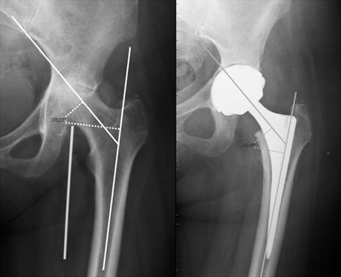
Fig. 6.2
Maintenance of the physiological angles
It is important to verify that the prosthesis does not impinge to the surrounding structures (a typical example is represented by the oversized prosthetic tibial plateau which impinges to the neighboring tendons causing inflammation of the anserine bursa) (Fig. 6.3).
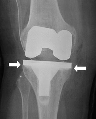
Fig. 6.3
Good positioning of prosthesis without impingement of tibial component with the surrounding structures
Subsequent radiological follow-up is useful to verify that the prosthesis maintains its position and functionality. Plain film still remains the first-choice technique, also in presence of the two main complications typical of prosthetic implantation: loosening (Fig. 6.4) (with possible periprosthetic fractures) and infection.
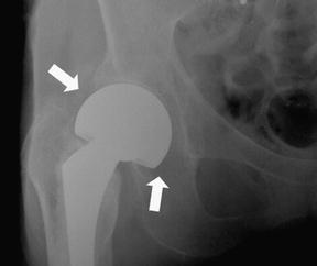
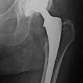
Fig. 6.4
Loosening and subsequent anteversion of the acetabular component
Hip—Besides being complete (acetabular or femoral) or only femoral, hip prostheses can be cemented or uncemented and the latter present a porous component that induces the periprosthetic growth of bone and the prosthetic adherence to the adjacent bone (Fig. 6.5). Hence, for the assessment of prosthetic loosening and possible presence of fracture, the medical specialists must bear in mind both the prosthetic characteristics and the original status of the patient. The prosthesis is cemented to guarantee a major longevity, but cement can induce osteolysis. Higher periprosthetic lucency around the bone–cement interface is observed in case of loosening of cemented prostheses (Fig. 6.6


Related posts:

Stay updated, free articles. Join our Telegram channel
Full access? Get Clinical Tree
