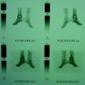
Fig. 7.1
Bone scintigraphy in posterior view. Aseptic loosening of the right hip prosthesis. The left prosthesis was painless. Significant uptake of the radiopharmaceutical can be seen around both the prostheses
The study of total knee replacement by bone scintigraphy is perhaps more problematic than that of hip prosthesis. In fact, it has been reported that 60 % of femoral and 90 % of tibial components show increased periprosthetic uptake more than 12 months after surgery [18, 19]. Serial bone scans performed in asymptomatic patients for a period of two years after implantation of total knee prostheses demonstrate that even if periprosthetic activity generally decreased over time, there was significant variability patient-to-patient [19]. These authors proposed the use of serial scans to actually asses the significance of increased periprosthetic uptake.
On this basis, the results obtained by further studies showing low accuracy of bone scan in assessing infection of total knee replacement are not surprizing [20, 21].
Bone scintigraphy can also be performed evaluating not only late images (reflecting bone osteometabolic activity) but also early phases of the scan (vascular and blood pool phases). A similar study is currently called 3-phase bone scan. This different approach does not seem to produce better results in diagnosing prosthetic infection, as reported in Table 7.1.
Table 7.1
Reliability of 3-phase bone scintigraphy in the assessment of prosthetic infection
Author and year | Type of prosthesis | N° of prostheses | Sensitivity (%) | Specificity (%) | Accuracy (%) |
|---|---|---|---|---|---|
Magnuson et al. [41] | Hip and knee | 49 | 100 | 18 | 53 |
Levitsky et al. [42] | Hip and knee | 72 | 30 | 86 | 68 |
Palestro et al. [20] | Knee | 41 | 67 | 76 | – |
Love et al. [21] | Hip and knee | 150 | 76 | 51 | 62* |
As can be seen, the clinical usefulness of bone scintigraphy in the evaluation of painful prosthetic joints can be considered low, due to the low values of its diagnostic accuracy. It does not seem possible with a single scan to differentiate aseptic loosening from either infection or normal post-operative appearance. For both cemented and porous-coated hip and knee replacements, bone scan is more useful in excluding prosthetic pathology when it is clearly negative. Due to its high negative predictive value, this scintigraphy can be used as a screening test or in association with other radionuclide studies.
7.2 Combined Bone/Gallium Scintigraphy
The property of gallium-67 of accumulating in both septic and aseptic inflammations has been known for about 40 years. Early investigations regarding the role of this radionuclide in musculo-skeletal infections showed variable values of accuracy. In 1979, Reing et al. carried out a study on 79 joint replacements, comparing bone and gallium-67 scintigraphies in diagnosing infection [22]. The sensitivity and specificity values obtained for bone and gallium-67 scans were 100, 15, and 95, 100 %, respectively. These findings suggested that gallium-67 imaging may significantly increase the accuracy of radionuclide diagnosis of infected joint replacement. Rushton et al. in a comparative study of bone and gallium-67 scans reported that gallium-67 accumulated in all the 13 infected prostheses they studied, whereas none of the 18 patients with aseptic loosening of prosthesis showed abnormal gallium-67 uptake (100 % accuracy) [23]. Other authors found 80 % accuracy values [13, 24], while Aliabadi et al. reported that gallium-67 scan is only 37 % sensitive, but 100 % specific [10]. In order to improve the accuracy of both bone and gallium-67 scans, the two studies are often interpreted together according to standardized criteria [25]. According to this approach, the scan is positive for infection when the spatial distribution of the two tracers is incongruent or when this distribution is spatially congruent but the gallium-67 uptake is higher than that found on the bone scan. The test is negative for infection when the gallium-67 scan is normal, independently from the bone scan results or when the spatial distribution of the two tracers is congruent, but the gallium-67 uptake is lower than that found on bone scan. Finally, the scans have to be considered dubious for infection when spatial distribution and intensity of uptake of the two radiotracers are congruent.
This combined interpretation, however, does not significantly increase the accuracy over either study alone. In fact, while Tehranzadeh et al. reported a 95 % accuracy for combined studies [26], the majority of the authors found less satisfactory results, as shown in Table 7.2.
In conclusion, on the basis of the above reported accuracy values, it can be affirmed that combined bone/gallium-67 imaging interpretation, produces only a slight accuracy improvement in comparison with bone scan alone.
7.3 Labeled Leukocyte Scintigraphy
From the physio-pathological point of view, indium-111-labeled leukocytes should be the most specific tracer for detecting infection, since they are always abundantly present in the histopathological specimen drawn at the site of prosthetic joint infection. Most of the early reported results, however, can be considered disappointing. A possible explanation of similar contradictory results may be found in the different methods used for image interpretation. All the methods shown in Table 7.3 are based on a comparison of intensities between periprosthetic region and another region used as reference point or merely on the presence of any periprosthetic activity. Labeled leukocytes accumulate in bone marrow, and even if bone marrow is more present in axial skeleton and proximal humeri and femurs, its distribution is characterized by significant inter-individual variability. Furthermore, orthopedic hardware can give rise to localized marrow expansion [27–29], and other systemic diseases can also cause generalized bone marrow expansion. Thus, similar conditions can make it difficult to correctly distinguish eventual alteration of marrow distribution from uptake really due to infection. Poor accuracy of above-mentioned results depends on the fact that the intensity of labeled leukocyte accumulation is not a useful criterion to obtain a correct diagnosis of infection.
Table 7.3
Diagnostic reliability of labeled leukocyte scintigraphy in the assessment of prosthetic infection
Author and year | Type of prosthesis | Criteria for classifying images as positive | Sensitivity (%) | Specificity (%) |
|---|---|---|---|---|
Pring et al. [46] | Hip and knee | Periprosthetic activity at least as intense as normal marrow | 100 | 89.5 |
Magnuson et al. [41] | Hip and knee | As above | 88 | 73 |
McKillop et al. [24] | Hip and knee | Any type of periprosthetic activity | 50 | 100 |
Wukich et al. [30] | Hip and knee | Focal increase of activity compared with adjacent bone marrow activity | 100 | 45 |
Johnson et al. [31] | Hip and knee | Any type of periprosthetic activity | 100 | 50 |
Palestro et al. [32] | Hip | Any type of periprosthetic activity, regardless of intensity | 100 | 23 |
Palestro et al. [32] | Hip | Periprosthetic activity more intense than the controlateral hip | 23 | 63 |
Palestro et al. [20] | Knee | Any type of periprosthetic activity, regardless of intensity | 89 | 50 |
Palestro et al. [20] | Knee | Periprosthetic activity more intense than the controlateral hip | 89 | 75 |
In order to improve accuracy of labeled leukocyte scans, combined leukocyte/bone scan was tested, assuming incongruent images with the two tracers as positivity criterion. As shown in Table 7.4, inconsistent results were obtained. In particular, some authors reported a significant increase in sensitivity (from 45 % with leukocyte scan to 85 % with leukocyte/bone imaging) against a slight drop in specificity (from 100 to 85 %) [30]. Further experiences gave similar results, showing higher specificity values of combined scans in comparison with leukocyte scan alone (95 vs. 50 %) and only small decrease of sensitivity (88 vs. 100 %) [31].
Table 7.4
Reliability of combined leukocyte/bone scintigraphy in the assessment of prosthetic infection
Author and year | Type of prosthesis | Sensitivity (%) | Specificity (%) |
|---|---|---|---|
Mulamba et al. [47] | Hip | 92 |