Rotator Cuff/Biceps Tendinosis
KEY FACTS
Terminology
Imaging
IMAGING
General Features
Ultrasonographic Findings
 Graded as mild, moderate, or severe based on degree of thickening, hypoechogenicity, and loss of normal fibrillar pattern
 Focal hypoechoic areas of proteoglycan deposition can mimic intrasubstance tears
 Nonintrasubstance tears are discernible through changes in tendon contour (contour retraction, loss of volume, fluid gap)
Imaging Recommendations
 Systematically examine tendons, taut and nontaut, in longitudinal and transverse planes with patient in sitting position
 Arm rotated into different positions to optimize visibility of each tendon in turn
 Important to always grade severity of tendinosis as mild, moderate, or severe for each tendon individually
DIFFERENTIAL DIAGNOSIS
Intrasubstance Tendon Tears
Rotator Cable
Rotator Cuff/Biceps Tendinosis


 indicative of tendinosis. A normal subscapularis tendon
  and biceps tendon
  are also shown.
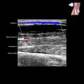
 indicative of moderate to severe tendinosis. A barely discernible fibrillary pattern
  is present. Note the hypoechoic area
  close to the insertion due to a combination of anisotropy and rotator cable effects.
 . Multiple, poorly demarcated, hypoechoic areas
  with loss of distinct fibrillary structure are present. Features are indicative of severe supraspinatus tendinosis.
 at proximal end of bicipital groove at “genu” between horizontal and vertical components. The more distal tendon is relatively normal
  . Tendinosis usually occurs at this location.