, Allan L. Brook2 and A. Orlando Ortiz3
(1)
Department of Radiology Quality/Safety Officer, Montefiore Medical Center, Bronx, NY, USA
(2)
Albert Einstein College of Medicine, Montefiore Medical Center, Bronx, NY, USA
(3)
Department of Radiology, Winthrop-University Hospital, Mineola, NY, USA
Keywords
Anatomy: sacrumSacroiliac jointBiopsy: paraspinal soft tissuesSacrumSacroiliac jointsImaging guidance: computed tomographyFluoroscopyPercutaneous sacral biopsyComplicationsSacral biopsy: contraindicationsIndicationsTechnique: coaxial techniqueLearning Objectives
- 1.
To understand the value and importance of radiologic anatomy as it pertains to sacral biopsy
- 2.
To review the indications and contraindications for biopsy of the sacrum and adjacent structures
- 3.
To learn the approaches and techniques for image-guided percutaneous sacral biopsy
7.1 Introduction
The sacrum, as a unit, is the largest “bone” within the spinal axis. This makes it a likely site for metastatic tumor involvement. Primary tumors can also occur within the sacrum. In terms of development of the spinal column, notochordal rests predispose the sacrum to the development of chordomas (the other common location for this lesion is the clivus at the skull base). Its articulation with the pelvic bones may also predispose to the sacrum, via the sacroiliac joints, to inflammatory and/or infectious processes. The location at the level of the pelvis may also predispose the sacrum to infectious and neoplastic processes that occur in this part of the body. Biomechanically, the sacrum can be affected by traumatic and weight-bearing loads that compromise its structural integrity. Therefore, with respect to size, embryologic developmental influences, location, and role in spine biomechanics, the sacrum can be affected by several pathologic conditions that may require image-guided percutaneous biopsy in order to establish a diagnosis and facilitate clinical management. The close proximity to the skin surface can create a misconception that a sacral biopsy is a straightforward procedure. The sacrum has an intricate anatomic relationship with the neuraxis, and an image-guided percutaneous sacral biopsy must always be performed using thorough preparation and meticulous technique.
7.2 Anatomic Considerations
The sacrum is a large complex triangular osseous structure that is located at the caudal aspect of the spinal axis between the lumbar spine and coccyx (Fig. 7.1) (Diel et al. 2001). The sacrum consists of five vertebrae, but unlike the more cephalad components of the spine, these vertebrae are fused anteriorly (vertebral bodies) and posteriorly (neural arches). A residual or persistent intervertebral disk is occasionally observed at the S1–S2 level. Additionally, transitional vertebral anatomy is not uncommon at the lumbosacral junction, with either a partially lumbarized S1 or a sacralized L5 vertebra present (Carrino et al. 2011). The superior aspect or S1 portion of the sacrum has more mass or bulk, and the sacrum thins or tapers down progressively from S1 to S5. The largest sacral vertebral body, S1, possesses a prominent anterior superior end plate, or promontory, with which to support the lumbar spine at the L5–S1 distal articulation. The spinous processes of the sacral neural arches are fused to form the median sacral crest. Unlike the other spine segments, the sacrum does not contain transverse processes. Instead, a continuous lateral osseous mass, the sacral ala, lies lateral to and is in continuity with the sacral vertebra and neural arches. A prominent marrow-containing space is present within the sacral ala, though the trabecular network is somewhat reduced as compared to other osseous structures within the spinal axis. The sacral ala articulates with the iliac bones at the sacroiliac joints. These large joints have a superior component that is fibrous and an inferior component that consists of a synovial joint space. The sacrum articulates caudally with the coccyx at the sacrococcygeal joint.


Fig. 7.1
Cross-sectional CT anatomy of the sacrum. Reformatted midline sagittal CT image (a) of the sacrum shows five fused sacral vertebrae bordered superiorly by the L5–S1 intervertebral disk (large arrow) and inferiorly by the sacrococcygeal joint (small arrow). The sacral hiatus (curved arrow) is seen at the S4–S5 level and defines the caudal extent of the sacral canal. Axial CT image (b) at the S1 level shows the sacral promontory (SP) and the sacral ala (SA). The sacral canal (large arrow) and S1 nerve root (small arrow) are seen at this level. The lumbosacral trunk is seen anterior to the sacral ala (oval). The upper sacroiliac joint (curved arrow) is seen between the sacral ala and the iliac bone. Axial CT image (c) at the S2 level shows the ventral (curved white arrow) and dorsal (small arrow) aspects of the neural foramina. The sacral canal (large arrow) is smaller in diameter at this level, while the sacroiliac joint (curved black arrow) is more prominent. The internal iliac artery (IA) is seen anterior to the sacral ala. Axial CT image (d) at the S3 level shows the smaller size of all sacral components, including the sacral canal (large arrow), as the sacrum tapers. The synovial portion of the sacroiliac joint (curved) arrow is seen at this level. Axial CT image (e) at S4 shows a small sacral vertebra and canal (arrow). The pyriformis (P) and gluteus (G) muscles form the anterior and lateral relations of the sacrum as does the rectosigmoid portion of the large intestine (curved arrow). Axial CT image (f) at the S5 level shows an aperture in the dorsal sacrum, the sacral hiatus, which is bordered by bony prominences, the sacral cornu
The lumbar spinal canal continues within the center of the sacrum as the sacral canal. The sacral canal courses through the center of the sacrum and contains the caudal aspect of the dural sac. The latter usually terminates at the S2 or S3 level. The remainder of the sacral canal contains epidural fat and an epidural venous plexus. The sacral canal terminates at the sacral hiatus, a small dorsal aperture at the caudal aspect of the sacrum which is bordered dorsally by two bony protuberances that are palpable on physical examination, the sacral cornu. The sacral canal communicates with the sacral foramina from S1 to S4. These sacral foramina are unique in that they possess ventral and dorsal apertures. The dorsal apertures of these foramina transmit sensory nerves which form a fibrous meshwork over the dorsum of the sacrum as they primarily innervate the sacroiliac joints. Motor nerves course through the anterior sacral foramina. The sacral plexus is formed by the ventral rami of the L4–S4 nerves and consists of a large upper band, or lumbosacral trunk, which derives neural contribution from L4, L5, and S1 and a small inferior band that includes branches from S2, S3, and S4.
The major ventral anatomic relations of the sacrum include the pelvis and pelvic contents. The presacral space contains fat. The rectum is located just anterior to this space. The iliac vessels, arteries, and veins are seen bilaterally anterior to the ventral sacral cortex at the level of the sacral ala. The lumbar plexus or lumbosacral trunk is also located adjacent to these structures. The sacral sympathetic plexus is located at the L5–S1 level between the common iliac vessels. The impar or sacrococcygeal ganglion is located anterior to the sacrococcygeal joint. The major lateral anatomic relations of the lower sacrum include the gluteal musculature and pyriformis muscles.
The critical anatomic sacral structures to be aware of for sacral biopsy include the sacral canal and foramina and the anterior sacral cortex.
7.3 Indications and Contraindications
Sacral biopsies are indicated to evaluate mass lesions within the sacrum and/or adjacent soft tissue structures (Table 7.1). These lesions may represent primary tumors in this location such as chordoma, giant cell tumor, or sarcoma (Figs. 7.2 and 7.3). More commonly, however, mass lesions of the sacrum are often due to metastatic disease from other primary sites (Fig. 7.4) (Rajeswaran et al. 2013). Infiltrative lesions of the sacrum may also require biopsy for definitive diagnosis. Infiltrative lesions may represent sacral involvement by multiple myeloma or lymphoma. When unilateral, sacral insufficiency fractures may show features that resemble an infiltrative lesion on MRI; this may require biopsy for clarification (Fig. 7.5) (Sudhir et al. 2016). Infection, or sacral osteomyelitis, is uncommon but may be seen in bedridden patients with sacral decubitus ulcers. Infection may also involve the sacroiliac joint, and this tends to present with unilateral involvement. Suspected infection in the sacrum, sacroiliac joint, or adjacent portions of the pelvis may require biopsy in order to confirm the diagnosis and identify the causative microorganism (such as Staphylococcus species).


Table 7.1
Indications/contraindications: image-guided percutaneous sacral biopsy
Indications |
Sacral mass |
Infiltrative sacral lesion |
Unable to distinguish unilateral sacral insufficiency fracture from infiltrative lesion |
Sacral osteomyelitis |
Contraindications |
Absolute |
Uncorrected coagulopathy |
Unable to obtain informed consent for the procedure |
Relative |
Uncooperative patient |
Unstable patient |

Fig. 7.2
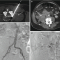
Chordoma. Contrast-enhanced T1-weighted sagittal image (a) shows large moderately enhancing septated mass within the distal sacrum (arrows); the mass projects ventrally. Axial CT image (b) shows the biopsy needle (arrow) within this expansile mass that is centered within the sacrum and erodes the surrounding bone

Fig. 7.3
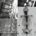
An 81-year-old male with low back and right leg pain. Fat-suppressed T2-weighted sagittal image (a) shows a marrow replacement process at S1 and S2 (arrows) and a ventral epidural soft tissue component (curved arrow). T1-weighted axial image (b) shows an infiltrative hypointense lesion within the right sacral ala (large arrow) and sacral vertebral body (small arrow) with a hypointense epidural soft tissue component (curved arrow). Single posterior projection (c) from a bone scan shows intense focal radionuclide uptake within the right side of the sacrum. Coronal reformatted fused image (d) from a PET-CT examination shows intense FDG uptake within the lesion (arrow). Axial CT image (e) obtained during the biopsy procedure shows a coaxial bone biopsy needle system with a guide cannula (large arrow) oriented in a standard short-axis oblique approach between the sacral foramina and right sacroiliac joint. The biopsy needle (short arrows) has been inserted into the right sacral ala. Subsequent pathologic evaluation of the biopsy specimens showed a high-grade fibroblastic sarcoma

Fig. 7.4
A 73-year-old female with prior medical history of melanoma, breast cancer and a recently detected lung mass presents with lesions in the sacrum and calvarium. Axial CT image (a) during biopsy procedure with skin grid in place shows a lytic soft tissue mass (arrow) that extends posteriorly. The arrow also indicates the desired trajectory for a posterior approach to this lesion. Axial CT image (b) shows a coaxial soft tissue biopsy system inserted with this oblique trajectory and with the guide cannula supported by gauze at the skin entry site (large arrow). The biopsy chamber (small arrows) of the cutting needle has been exposed within the substance of the lesion. The pathology showed metastatic lung adenocarcinoma

Fig. 7.5
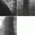
A 47-year-old female with gait disturbance. Posterior frontal projection (a) from bone scan shows focal spotty radionuclide uptake within the lower sacrum (arrow). T1-weighted sagittal image (b) shows focal hypointensity at S3 and S4 (arrow) with corresponding hyperintensity (arrow) on the T2-weighted sagittal image (c). Fat-suppressed contrast-enhanced T1-weighted coronal images (d) show focal enhancement within the lower sacrum as well as the presence of a fracture line (arrows). In retrospect, the irregular linear pattern of the sacral insufficiency fracture line is shown on the bone scan
Sacral biopsies are strictly contraindicated in patients with uncorrected coagulopathy (Table 7.1). The procedure cannot be performed without informed or administrative consent. Relative contraindications to sacral biopsy include an uncooperative patient or an unstable patient. With respect to metastatic tumor, in situations where neoplastic lesions are present in both the sacrum and iliac bone, the operator may consider performing the iliac bone biopsy instead (Fig. 7.6).


Fig. 7.6
A 51-year-old female with lower low back pain. Axial CT image obtained during biopsy procedure shows sampling of a sclerotic lesion within the iliac crest (large arrow) with a coaxial bone biopsy needle system (short arrow). This iliac crest was biopsied instead of the sclerotic lesion within the adjacent sacral ala in this patient with breast metastases
7.4 Risks and Complications Associated with Sacral Biopsy and How to Minimize Them
A sacral biopsy is an invasive procedure, and the possibility of a complication, though uncommon, should not be ignored (Table 7.2). Prerequisites that will help to minimize the complication rate when performing image-guided percutaneous spine biopsy procedures such as sacral biopsy are based upon fundamental surgical principles and consist of four major concepts. First, patient selection is important. The procedure should be indicated, and the contraindications to the procedure should be adhered to. If there are relative contraindications to the procedure, then they should be reconciled, with patient safety at the forefront of this decision, prior to moving forward with the procedure. Second, patient preparation must take place, including a review of the patient’s medical record, an understanding of the patient’s medical condition(s), and their ability and willingness to have the procedure. Medications and medical allergies must be known ahead of time and the appropriate steps taken to temporarily adjust medication regimens when necessary. Third, operator preparation is essential to a successful and safe procedure. This concept not only reflects the operators training, abilities, and knowledge with respect to image-guided percutaneous biopsy techniques (including a sound understanding of the imaging modalities, tools, and techniques for performing these procedures) but also emphasizes the basic questions of should I perform this biopsy, when do I perform this biopsy, and how should I perform this biopsy (Carberry et al. 2016)? At the clinical level, thorough preparation should reflect careful patient evaluation, image review, and communication with the referring clinician. Fourth, the value of an organized team approach toward performing the procedure is a major determinant in the success of the procedure and in a safe patient outcome. This includes all aspects of the procedure from patient intake, evaluation, preparation, procedure (inclusive of all appropriate procedural suite protocols such as sterile technique, patient and procedure verification), post-procedure care, and patient follow-up. The premise that everyone’s role, including the patient and their family, in patient safety is equally important, and the actual practice of this credo improves the likelihood of a successful outcome.
Table 7.2
Sacral spine biopsy procedure risks and complications
Hemorrhage |
Superficial – at puncture site |
Deep – potential pelvic hematoma formation |
Needle injury |
Artery or vein puncture |
Spinal canal or foramen breach |
Sacral nerve root or lumbosacral trunk puncture |
Other |
Nondiagnostic biopsy |
Wrong side biopsied |
Infection (cellulitis, osteomyelitis) |
Tumor seeding along the biopsy tract
Stay updated, free articles. Join our Telegram channel
Full access? Get Clinical Tree
 Get Clinical Tree app for offline access

|


