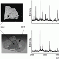
Fig. 1
Mousetrap design used for the freeze fracture system. The sample is trapped between two metal plates connected by a hinge, which is sprung open when triggered. Reproduced from [5] with permission from John Wiley and Sons
5.
Transfer sample to a liquid nitrogen flask containing the mousetrap device where the sample is mechanically fixed in place.
6.
Transfer the sample to the instrument through the use of a glove box purged with argon gas to prevent frosting.
7.
Sample is mounted directly onto a precooled sample insertion stage and transferred into the preparatory chamber.
8.
Sample is fractured in the instrument at 168 K to minimize deposition of water. The trap is sprung by a transfer arm revealing cryogenically preserved cells (see Note 5 ).
9.
Sample is transferred to a cold stage in the analysis chamber and held below 150 K throughout analysis (see Note 6 ).
3.3 Frozen Hydrate [13]
1.
To prepare cells for analysis, add the desired amount of cells to a petri dish containing clean and dry silicon shards and allow growth in incubator until optimal coverage is obtained, usually about 24 h (see Note 7 ).
2.
Warm 0.15 M ammonium formate solution (pH ~7.3) to 37 °C.
3.
Cool the sample holder in liquid nitrogen after blowing it dry with nitrogen gas. Allow the holder to cool in the liquid nitrogen and do not add the sample until the boiling has stopped. Keep the holder submerged in liquid nitrogen for the duration of the sample preparation (see Note 8 ).
4.
Wash the cells three times for ~5 s, in three beakers of the 0.15 M ammonium formate solution, to remove residual salts present from the cell media for a total of nine washes (see Note 4 ).
5.
Dry the shards with a very gentle stream of nitrogen. If the nitrogen pressure is too great, streaking of the cells will occur. Also, be sure to dry the tweezers prior to freezing the sample (see Note 9 ).
6.
Quickly plunge freeze the shard with cell growth into liquid propane for about 3–4 s before transferring to the liquid nitrogen-covered sample holder. If transfer times are too slow, the sample will warm above ~165 K and the cells will rupture.
7.
Transfer into instrument for analysis, again, minimizing the amount of time that the cells are exposed to room temperature. The instrument should be operated with a precooled stage.
3.4 Chemical Fixation with Glutaraldehyde [6]
1.
Seed the hTERT-BJ1 onto the clean silicon shards and allow growth in an incubator to occur for up to 2 days.
2.
Wash the cells with phosphate-buffered saline (PBS).
3.
Fix the cells with 2.5 % glutaraldehyde for 15 min in 37 °C.
4.
Wash away excess GA with PBS.
5.
Gently dry with nitrogen.
6.
Plunge freeze in liquid propane and isopentane (3:1). This allows for a high cooling rate, reducing water crystallization.
7.
Allow to freeze-dry overnight at −80 °C and 10−6 mbar.
8.
Allow to warm by 10 °C/h to 30 °C.
9.
If freeze-drying did not occur in the SIMS instrument, transfer sample into the instrument and complete analysis.
3.5 Chemical Fixation with Paraformalin/Formaldehyde [7]
1.
To prepare cells for analysis, add the desired amount of cells to a 24-well plate containing clean and dry poly-l-lysine-coated silicon shards and allow growth in incubator for 24 h until optimal coverage is obtained (see Note 3 ).
2.
Aspirate the media.
3.
Wash with phosphate-buffered saline (PBS) three times.
4.
Incubate the cells with 4 % formalin for 15 min at 4 °C.
5.
Wash with PBS five times and then wash with water three times.
6.
Return to PBS for storage for several hours.
7.
Wash with 0.15 M ammonium formate for 1 min and allow to dry.
8.
Transfer into the SIMS instrument and begin analysis.
3.6 Chemical Fixation with Trehalose [16]
1.
Cells were allowed to grow for several days prior to fixation.
2.
Incubate cells in 50 mM trehalose for several hours.
3.
Rinse for ~5 s with PBS buffer containing 50 mM trehalose and 10–15 % glycerol by weight. The addition of glycerol ensures stronger adhesion of the cells to the substrate.
4.
Place a 200-mesh grid, followed by a thin substrate on top of the sample, creating a sandwich, and freeze in liquid nitrogen. Since trehalose acts similarly to a cryopreservant, flash freezing is not necessary in this protocol.
5.
Place samples under vacuum and leave at 10−2–10−7 mbar overnight (at least 15 h) to allow freeze-drying to occur.
Stay updated, free articles. Join our Telegram channel

Full access? Get Clinical Tree


