This appendix is provided as a guide to the technical aspects of various imaging procedures. Some of the less common procedures have not been included, and the procedures described herein may need to be adjusted, depending on the equipment available and user preferences. The protocols for positron emission tomography (PET) examinations are at the end of this appendix. Each nuclear medicine laboratory should have a standardized procedures manual; this appendix may be used as a beginning point for the development of such a manual. The reader is also referred to manufacturers’ recommendations and the procedure standards in the Guidance section under the Quality and Practice heading of the Society of Nuclear Medicine website ( http://www.snm.org ). Suggested administered activities for pediatric and adolescent examinations are given in Appendix D .
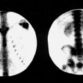
Brain Death or Cerebral Blood Flow Scan
Procedure imaging time
20 to 30 minutes
Radiopharmaceutical
Technetium-99m ( 99m Tc) or diethylenetriamine pentaacetic acid (DTPA) for planar imaging. Brain-specific single-photon emission computed tomography (SPECT) perfusion agents, such as 99m Tc-hexamethylpropyleneamine oxime (HMPAO) and 99m Tc-ethyl cysteinate dimer (ECD), also called 99m Tc-bicisate, can also be used, but there is no clear evidence that they are more accurate, although they are less dependent on an excellent bolus injection.
Method of administration
Bolus IV injection for any radiopharmaceutical used
Normal adult administered activity
99m Tc-DTPA 15 to 30 mCi (555 MBq to 1.11 GBq)
99m Tc-HMPAO and ECD 10–20 mCi (370–740 MBq)
Injection-to-imaging time
Immediate with delays as warranted
Conflicting examinations and medications
None
Patient preparation
None necessary, although some institutions put a rubber band or tourniquet around the head just above ears to help diminish scalp blood flow. This should not be done in patients with a history of head trauma. Patient should be normally ventilated.
Technique
Collimator
High-resolution or ultrahigh-resolution; field of view (FOV) should include from the level of the common carotids to the skull vertex.
Dynamic flow imaging time
Blood flow images: 1 to 3 seconds/frame for at least 60 seconds. Flow images should start before the arrival of the bolus in the neck.
Routine views
Immediate blood pool anterior and anterior image at 5 minutes each. Many institutions also obtain posterior and both lateral views. 128 × 128 matrix. Note: If brain-specific images are obtained, initial images as described previously are obtained as well as planar and SPECT images obtained after 20 minutes.
Patient positioning
Sitting or supine
Photopeak selection
140-keV (15% to 20% window)
Dosimetry: rads/mCi (mGy/MBq) of administered activity
Effective dose
0.0122 (0.0033)
HMPAO (also called Ceretec or exametazime )
Effective dose
0.0363 (0.0098)
Effective dose
0.0207 (0.0056)
SPECT Brain Perfusion Imaging
Procedure imaging time
30 to 60 minutes
Instrumentation
SPECT camera
Radiopharmaceutical
99m Tc-HMPAO (exametazime, unstabilized or stabilized), 99m Tc-ECD.
For unstabilized 99m Tc-HMPAO, inject no sooner than 10 minutes after preparation and not more than 30 minutes after preparation. For seizure disorders, inject within 1 minute after reconstitution. For stabilized 99m Tc-HMPAO, inject no sooner than 10 minutes after preparation and no more than 4 hours after preparation. For 99m Tc-ECD, inject no sooner than 10 minutes after preparation and no more than 4 hours after preparation.
Method of administration
Place patient in a quiet, dimly lit room and instruct him or her to keep eyes and ears open. The patient should be seated or reclining comfortably. IV access should be placed at least 10 minutes before injection. The patient should not speak or read, and there should be little or no interaction before, during, or up to 5 minutes after injection.
Normal administered activity
15 to 30 mCi (555 MBq to 1.11 GBq), children 0.3 mCi/kg (11.1 MBq/kg). Minimum dose 5 mCi (185 MBq).
Injection-to-imaging time
90 minutes or later for stabilized or unstabilized 99m Tc-HMPAO, although images obtained after 40 minutes will be interpretable; 45-minute delay for 99m Tc-ECD, although images obtained after 20 minutes will be interpretable. If possible, all imaging should be obtained within 4 hours of injection.
Conflicting examinations and medications
None
Patient preparation
Patient should be instructed, if possible, to avoid caffeine, alcohol, or other drugs known to affect cerebral blood flow. If sedation is required, it should be given after the injection and after the radiopharmaceutical uptake period. Patient should void before study for maximum comfort and to prevent scan interruption.
Technique
Collimator
Low-energy, high-resolution or ultrahigh-resolution, or fan beam; all-purpose
Acquisition
128 × 128 or greater acquisition matrix; 3-degree or better angular sampling. Acquisition pixel size should be one-third to one-half of the expected resolution. Low-pass Butterworth filters are preferred for processing in all three dimensions. Attenuation correction should be performed.
Routine views
360-degree arc of rotation single head camera; however, multiple head detectors may produce better images.
Patient positioning
Supine
Photopeak selection
140-keV 99m Tc (20% window)
Dosimetry: rads/mCi (mGy/MBq) administered
HMPAO (also called Ceretec or exametazime )
Effective dose
0.0363 (0.0098)
Effective dose
0.0207 (0.0056)
Comments
Vasodilatory challenge with acetazolamide (Diamox) may be ordered for evaluation of cerebrovascular reserve in transient ischemic attack, completed stroke, or vascular anomalies. Known sulfa allergy is a contraindication, and the procedure is usually avoided within the first 3 days after an acute stroke. The challenge study is usually done first, and, if normal, the baseline study may be omitted. The dosage is 1000 mg in 10 mL sterile water by slow IV push (over 2 minutes) and 14 mg/kg for children. Wait 10 to 20 minutes before injecting tracer. The patient should void immediately before acquisition.
Cisternogram
Procedure imaging time
30 minutes for each set of images
Instrumentation
Planar gamma camera
Radiopharmaceutical
Indium-111 ( 111 In)-DTPA pyrogen free
Method of administration
Spinal subarachnoid space injection
Normal adult administered activity
0.5 mCi (18.5 MBq)
Injection-to-imaging time
2 hours, 6 hours, 24 hours, 48 hours, and 72 hours (as needed).
Conflicting examination and medications
Acetazolamide (Diamox) can cause false-positive results.
Patient preparation
If the clinical diagnosis is cerebrospinal fluid (CSF) rhinorrhea or otorrhea, the patient’s nose or ears should be packed with pledgets before injection for later counting.
Technique
Collimator
Medium energy parallel-hole
Counts
- 1.
50- to 100-k counts for 111 In.
- 2.
Cobalt ( 57 Co) for 50-k counts transmission scan (if useful for anatomic definition).
- 1.
Routine views
- 1.
Anterior transmission scan: position patient’s head between 57 Co sheet source and collimator surface. Peak in 57 Co by after photopeak determination. Set intensity, but collect only 50-k counts. Do not advance film or image. Remove sheet source from behind patient. Peak detector for 111 In. Collect 100-k counts.
- 2.
Lateral transmission scan.
- 3.
Anterior head.
- 4.
Lateral head (same lateral as transmission scan).
- 1.
Patient positioning
Supine. If a significant CSF leak is suspected in a specific area, the patient may be positioned with that portion dependent.
Photopeak selection
57 Co (for transmission images) 122-keV; 111 In-DTPA 173-keV (20% window).
Dosimetry: rads/mCi (mGy/MBq) of administered activity
Effective dose
0.197 (0.0533)
Comments
For CSF rhinorrhea or otorrhea, count all pledget samples in well counter after removal from nose and ears. Note: Remove the pledgets and place each in a separate counting vial at time of removal, labeling each vial with its location.
Thyroid Scan ( 99m Tc-Pertechnetate)
Procedure imaging time
15 minutes
Radiopharmaceutical
99m Tc-sodium pertechnetate
Method of administration
IV injection
Normal administered activity
Adult, 2 to 10 mCi (74 to 370 MBq). For children, 0.14 mCi/kg (5 MBq/kg), minimum of 0.27 mCi (10 MBq).
Injection-to-imaging time
15 to 30 minutes
Conflicting examinations and medications
None
Patient preparation
None
Technique
Collimator
Low-energy parallel and pinhole
Counts
50-k counts per image or 5 minutes (whichever is sooner). 128 × 128 matrix.
Patient positioning
- 1.
Supine.
- 2.
Extend neck forward by placing a positioning sponge under back of neck.
- 1.
Routine views
- 1.
Anterior view of the thyroid to include salivary glands, using parallel collimator.
- 2.
Pinhole views of thyroid only, in anterior and both anterior oblique positions (positioned so that the thyroid gland fills two thirds of the FOV).
- 1.
Photopeak selection
140-keV (20% window)
Dosimetry: rads/mCi (mGy/MBq) of administered activity
Effective dose
0.0585 (0.0158)
Comments
Remind the patient not to swallow during imaging. Drinking water followed by reimaging is sometimes useful to eliminate confusing esophageal activity.
Thyroid Scan and Uptake (Iodine-123)
Procedure imaging time
1 hour
Radiopharmaceutical
Iodine-123 ( 123 I) sodium iodide
Method of administration
Oral
Normal administered activity
200 to 400 µCi (7.4 to 14.8 MBq). For 5-year-old child, 3 to 10 µCi/kg (0.1 to 0.3 MBq/kg).
Administration-to-imaging time
3 to 24 hours
Conflicting examinations and medications
- 1.
Radiographic procedures using IV iodine contrast media (e.g., IV pyelogram, computed tomography [CT] scan with contrast).
- 2.
Other radiographic procedures using iodine contrast media (e.g., myelogram, oral cholecystogram).
- 3.
Exogenous T 3 or T 4 (liothyronine, levothyroxine).
- 4.
Thyroid-blocking agents such as propylthiouracil, perchlorate, and methimazole.
- 5.
Oral iodides in medications containing iodine (e.g., kelp preparations, vitamins, Lugol solution).
- 6.
If necessary, do a pertechnetate ( 99m TcO 4 − ) scan.
- 1.
Patient preparation
Scanning dose to be administered 3 to 24 hours before scanning. Patient should take nothing by mouth (NPO) overnight before examination. If patient is pregnant or lactating, consider using 99m TcO 4 − .
Technique
Collimator
Pinhole
Counts
50- to 100-k counts per image or 10 minutes/image
Routine views
Anterior, right, and left anterior oblique
Patient positioning
Supine, neck extended
Photopeak selection
159-keV (20% window)
Dosimetry: rads/mCi (mGy/MBq) of administered activity
Oral administration, medium uptake
Effective dose
0.788 (0.213) 35% uptake
0.0366 (0.099) 15% uptake
0.0333 (0.009) 0% uptake
Comments
- 1.
Iodine uptake is normally measured at 24 hours, although it may be measured at 6 hours, if appropriate. It is measured with a sodium iodide probe.
- 2.
Patient’s thyroid should be palpated by the physician, especially if the patient presents with nodular disease.
- 1.
Thyroid Cancer Scan
Procedure imaging time
1 to 2 hours
Radiopharmaceutical
131 I-sodium iodide or 123 I-sodium iodide
Method of administration
Oral
Normal adult administered activity
1 to 5 mCi (37 to 185 MBq) 131 I-sodium iodide
1 to 2 mCi (37 to 74 MBq) 123 I-sodium iodide
10 to 20 mCi (370 to 740 MBq) 99m Tc-sestamibi
Injection-to-imaging time
72 hours (96 hours, if needed) 131 I-sodium iodide
24 hours for 123 I-sodium iodide
15 minutes for 99m Tc-sestamibi
Conflicting examinations and medications
Iodine-containing medications and contrast agents
Patient preparation
For radioiodine, 2 weeks off T 3 replacement or 4 to 6 weeks off T 4 replacement. In some patients, the use of rTSH (thyrogen) may be useful to supplement or avoid thyroid hormone withdrawal. Some institutions use a low-iodine diet 3 to 10 days before administration of tracer.
Technique
Whole-body scan or spot views of head, neck, chest, and other clinically suspect areas.
Collimator
Medium- or high-energy for 131 I-sodium iodide or low-energy for 123 I-sodium iodide or 99m Tc
Counts
200-k counts or 10-minute spot views
Routine views
Anterior and posterior whole-body views
Patient positioning
Supine
Photopeak selection
364-keV (20% window) for 131 I-sodium iodide or 159-keV (20% window) for 123 I-sodium iodide.
Absorbed dose with thyroid removed or ablated.
Dosimetry: rads/mCi (mGy/MBq) of administered activity
Oral administration, thyroid (removed) uptake 0%
123 I
Effective dose
0.0341 (0.00923)
131 I
Effective dose
0.0154 (0.00416)
Effective dose
0.0244 (0.0066)
Comments
- 1.
This scan for metastatic disease is typically done after ablation of normal/residual thyroid tissue.
- 2.
Serum thyroid-stimulating hormone (TSH) levels should be above 40 mU/mL before start.
- 3.
Scanning can also be done 7 to 10 days after a cancer therapy treatment with 131 I.
- 4.
Scanning with 123 I may prevent stunning of thyroid remnant or metastases.
- 5.
Occasionally, scans are done by using 18 F-FDG, 99m Tc-sestamibi, or thallium-201 ( 201 Tl) chloride to locate nonfunctioning (nonradioiodine-avid) metastases.
- 1.
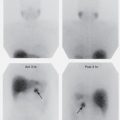
Parathyroid Scan
Procedure imaging time
2 hours
Radiopharmaceutical
99m Tc-sestamibi
Method of administration
IV administration
Normal adult administered activity
Planar 20 mCi (740 MBq); SPECT 30 mCi (1.1 GBq)
Injection-to-imaging time
5 minutes
Conflicting examinations and medications
None
Patient preparation
None
Technique
Collimator
Low-energy, high-resolution, or pinhole
Counts/time
Acquire image for 10 minutes, and if digital acquisition, use a 128 × 128 or larger matrix.
Routine views
Planar. Anterior images of the neck at 5, 20, and 120 minutes after injection. A single anterior large-FOV image should also be obtained that includes the mediastinum.
SPECT. Begin after 10 minute planar image. 180 degrees clockwise, 64 steps, 15 sec per step.
Patient positioning
Supine
Photopeak selection
140-keV (20% window)
Dosimetry: rads/mCi (mGy/MBq) of administered activity
Effective dose
0.0244 (0.0066)
Rest Gated Equilibrium Ventriculography
(Stress study and computer operation vary widely and are not presented here.)
Procedure imaging time
30 minutes
Radiopharmaceutical
99m Tc-labeled red blood cells (RBCs). See RBC labeling procedures at the end of this appendix. The modified in vivo method is suitable for this examination, although some laboratories use commercial in vitro methods such as Ultratag for convenience.
Method of administration
IV injection
Normal adult administered activity
15 to 30 mCi (555 MBq to 1110 MBq)
Injection-to-imaging time
Immediate
Conflicting examinations and medications
None
Patient preparation
Fasting for 3 to 4 hours before the study is preferred
Technique
Collimator
Low-energy, all-purpose, or high-resolution parallel-hole
Counts
3 to 7 million counts with a minimum of 16 and preferably 32 to 64 frames per second
Patient positioning
Supine or upright
Photopeak selection
140-keV (20% window)
Dosimetry: rads/mCi (mGy/MBq) of administered activity
Effective dose
0.0407 (0.011)
Comments
In vivo RBC labeling.
- 1.
Dilute a vial of cold pyrophosphate with 1 to 3 mL of sterile saline (not bacteriostatic). Shake the mixture, and let it stand for 5 minutes. Without injecting air into the vial, withdraw the contents into a 3-mL syringe, avoiding inclusion of an air bubble.
- 2.
Inject patient with cold pyrophosphate (0.8 to 1 mg stannous chloride).
- 3.
After 20 minutes, inject the radiopharmaceutical.
- 4.
Connect electrocardiogram leads to patient 5 to 10 cm below the axilla bilaterally. Remember to abrade the skin well enough so that the leads have good contact.
- 5.
Place the patient in the supine position on an imaging table with left side toward the camera.
- 1.
SPECT Myocardial Perfusion Imaging
Procedure imaging time
30 minutes for each set of images
Instrumentation
SPECT camera
Radiopharmaceutical
99m Tc-sestamibi, 201 T1-chloride, or 99m Tc-tetrofosmin
Method of administration
IV injection
Normal adult administered activity (same day stress-rest)
99m Tc-sestamibi
Rest 8–12 mCi (296–444 MBq)
Stress 24–36 mCi (888–1332 MBq)
201 T1-chloride
Stress or rest 2–4 mCi (111–185 MBq)
Stress/rest 3 mCi (111MBq) followed by 1 mCi (185 MBq) before 3–4 hour redistribution image
99m Tc-tetrofosmin
Rest 8–12 mCi (296–444 MBq)
Stress 24–36 mCi (888–1332 MBq)
Injection-to-imaging time
For post-stress images, immediate for thallium, approximately 15 to 20 minutes for tetrofosmin or sestamibi. Because there is minimal redistribution with technetium agents, longer delays up to 2 hours can be used when needed.
Conflicting examinations and medications
Discontinue calcium antagonists, β-blockers, and nitrates, if possible.
With thallium, increased myocardial uptake has been reported with dipyridamole, furosemide, isoproterenol sodium bicarbonate (IV), and dexamethasone; decreased myocardial uptake with propranolol, digitalis, doxorubicin, phenytoin (Dilantin), lidocaine, and minoxidil.
Patient preparation
NPO for 4 hours; exercise, if required. In patients with severe coronary disease, it may be advisable to administer nitroglycerin sublingually about 3 minutes before rest injection of the radiopharmaceutical.
Technique
Collimator
Low-energy, all-purpose
Counts and time
30 to 32 stops for 40 seconds each for 201 Tl and 25 seconds for 99m Tc sestamibi
Routine views
180- or 360-degree arc of rotation; 180 degrees is preferred from right anterior oblique to left posterior oblique. Either step and shoot acquisition with 32 or 64 stops separated by 3 to 6 degrees or continuous acquisition may be used. The duration for each stop varies but is generally 40 seconds per image for thallium and 25 seconds for technetium radiopharmaceuticals.
Patient positioning
Supine, left arm overhead
Photopeak technetium
85-keV (15% window) and possibly a second window 135- to 160-keV for thallium and 140-keV (20% window) for technetium.
Dosimetry: rads/mCi (mGy/MBq) of administered activity
Effective dose
0.3789 (0.102)
Effective dose
0.0244 (0.00661) rest
0.023 (0.00629) stress
Effective dose
0.0228 (0.00615) rest
0.0210 (0.00567) stress
Comments
- 1.
Process for short- and long-axis views.
- 2.
Parametric images such as “bull’s eye” maps can be used to map and semiquantitate regional perfusion compared to pooled normal gender-specific data.
- 3.
Use 64 × 64 matrix, Butterworth filter, 0.4 cutoff.
- 4.
Attenuation correction significantly reduces artifacts.
- 1.
Exercise Protocol Using a Bruce Multistage or Modified Bruce Treadmill Exercise Protocol
Patient preparation
Initial imaging
For 99m Tc-sestamibi, SPECT is performed a minimum of 15 to 20 minutes after exercise injection, 45 to 60 minutes for rest, and 60 minutes for pharmacologic stress.
For 99m Tc-tetrofosmin, minimum delays of 10 to 15 minutes for exercise, 30 to 45 minutes for rest, and 45 minutes for pharmacologic stress are recommended.
After-exercise instructions
Only light food intake; minimal physical exertion
Redistribution imaging
3 to 4 hours after injection
Contraindications
Unstable angina with recent (less than 48 hours) angina or congestive heart failure, documented acute myocardial infarction within 2 to 4 days of testing, uncontrolled systemic (systolic greater than 220 mm Hg, diastolic greater than 120 mm Hg) or pulmonary hypertension, untreated life-threatening arrhythmias, uncompensated congestive heart failure, advanced atrioventricular block (without a pacemaker), acute myocarditis, acute pericarditis, severe mitral or aortic stenosis, severe obstructive cardiomyopathy, and acute systemic illness. Relative contraindications to exercise stress include conditions that may interfere with exercise such as neuralgic, arthritic, or orthopedic conditions or severe pulmonary or peripheral vascular disease.
Dipyridamole Pharmacologic Stress Procedure
Patient preparation
NPO 4 to 6 hours; withhold caffeine-containing beverages for at least 12 hours and preferably 24 hours.
Drug administered
IV infusion of dipyridamole in antecubital vein with patient supine; rate, 0.5 mg/kg over 4 minutes in 20 to 40 mL of normal saline.
Radiopharmaceutical administered
IV administration of any of the radiopharmaceuticals listed above 3 minutes after dipyridamole infusion, with patient supine or upright.
Imaging
Begin SPECT imaging 3 to 4 minutes after thallium injection; repeat in 3 to 4 hours. For technetium radiopharmaceuticals, postinjection imaging time is not as critical and may be done at 30 to 60 minutes.
Comments
Side effects may be reversed by IV administration of 100 to 200 mg of aminophylline over 1 minute. No caffeine or theophylline for 12 hours before procedure.
Adenosine Pharmacologic Stress Procedure
Patient preparation
Contraindicated in patients with second- or third-degree atrioventricular block, sinus node disease, or asthma. Withhold dipyridamole for 12 to 24 hours before adenosine.
Drug administered
Adenosine, 140 mcg/kg per minute peripheral IV infusion over 6 minutes (total dose, 0.84 mg/kg) or 50 mcg/kg per minute increased to 75, 100, and 140 mcg/kg per minute each minute to 7 minutes.
Radiopharmaceutical administered
Administered at the midpoint (3 minutes) of the infusion.
Imaging
Post-stress imaging is performed as appropriate for the radiopharmaceutical employed.
Comments
Side effects of hypertension, flushing, chest discomfort, dyspnea, headache, dizziness, or gastrointestinal discomfort may occur and usually resolve quickly, although theophylline (50 to 125 mg slow IV injection) may be necessary in rare cases.
Regadenoson Pharmacologic Stress Procedure
Patient preparation
Contraindicated in patients with second- or third-degree atrioventricular block, sinus node dysfunction without a functioning pacemaker, systolic blood pressure less than 90 mm Hg. Regadenoson should be used with caution in patients with known or reactive airways disease or profound bradycardia (HR less than 40 bpm).
Drug administered
Regadenoson (5 mL containing 0.4 mg of regadenoson) is administered as a rapid (approximately 10 seconds) injection into a peripheral vein using a 22-gauge or larger catheter or needle. This is immediately followed by a 5 mL saline flush.
Radiopharmaceutical administered
Administered 10–20 seconds after the saline flush
Imaging
Imaging is performed as appropriate for the radiopharmaceutical employed.
Comments
Reversal of the effects of Regadenoson with aminophylline should be considered in the presence of severe hypotension (systolic BP less than 80 mm Hg), development of heart block, onset of bronchoconstrictive symptoms, or persistent chest pain or significant ST depression.
Pulmonary Ventilation Scan (Xenon)
Procedure imaging time
5 minutes
Instrumentation
Large-FOV camera, if available
Radiopharmaceutical
Xenon-133 ( 133 Xe)
Method of administration
Gas is inspired through an enclosed ventilation system with appropriate mouthpiece or face mask.
Normal administered activity
Adults, 5 to 20 mCi (200 to 740 MBq). (If done after perfusion scan, dose may have to be 20 mCi [740 MBq]).
Children, 0.3 mCi/kg (10 to 12 MBq/kg) with a minimum of 3 mCi (100 to 120 MBq).
Conflicting examination and medications
None
Patient preparation
None
Technique
Collimator
Planar: Low-energy, all-purpose, parallel-hole
Counts
10-second image for inspiration; 30 seconds for all other images; matrix 128 × 128
Routine views
All views are performed in the posterior position unless otherwise specified by the supervising physician.
- 1.
Begin 133 Xe administration and obtain 10-second inspiration image (~1000-k counts).
- 2.
Record three equilibrium images 30 seconds each.
- 3.
Exhale 133 Xe and record 30-second images until the bulk of the gas has left the lungs.
- 1.
Patient positioning
Sitting, preferably. Supine is also acceptable.
Photopeak selection
81-keV (25% window)
Dosimetry: rads/mCi (mGy/MBq) of administered activity (5 minutes rebreathing)
Effective dose
0.0044 (0.0012)
Comments
An exhaust system or xenon trap should be available for expired xenon or the exhaled xenon exhausted to the outside atmosphere. Room may be negative pressure.
Pulmonary Ventilation Scan (DTPA Aerosol)
Procedure imaging time
15 minutes
Instrumentation
Large-FOV camera, if available
SPECT/CT, if appropriate
Radiopharmaceutical
99m Tc-diethylenetriamine pentaacetic acid (DTPA) aerosol
Method of administration
Nebulizer connected to a mouthpiece and oxygen (typically 10 L/min). Patient performing normal tidal breathing in the upright position, if possible. Avoid deep or tubulent breathing to lessen hot spots and central deposition.
Normal adult administered activity
25 to 35 mCi (900 to 1300 MBq) in the nebulizer from which the patient receives about 0.54 to 1.1 mCi (20 to 40 MBq). The lower values used if to be followed by 99m Tc-macroaggregated albumin (MAA) perfusion scan.
Conflicting examination and medications
None
Patient preparation
Have patient practice breathing through nebulizer mouthpiece. The nose should be occluded.
Technique
Collimator
Planar: Low-energy, all-purpose, parallel-hole
SPECT/CT: High resolution
Photopeak selection
140-keV (20% window)
Counts
200K counts for first view (anterior/posterior), then all subsequent views for same amount of time or 3 minutes/view for all views, whichever is shorter. Note time required to take first posterior image.
Routine views
Anterior, posterior, and four obliques
Patient positioning
Sitting, preferably. Supine is also acceptable.
Dosimetry: rads/mCi (mGy/MBq) of administered activity
Effective dose
0.0226 (0.0061)
Comments
Usually performed before the 99m Tc-MAA perfusion scan.
Pulmonary Perfusion Scan
Procedure imaging time
30 minutes
Radiopharmaceutical
99m Tc-MAA, about 300,000 (200,000 to 700,000) particles; however, this should be reduced in children, in adults with known right-to-left shunts, and pulmonary hypertension (see comments below).
Method of administration
Before injection the patient should, if possible, cough and take several deep breaths. Invert syringe immediately before injection to resuspend particles. With the patient supine, or as close to supine as possible, begin slow IV injection in antecubital vein during three to five respiratory cycles.
Normal administered activity
Adult 1.1 to 4.1 mCi (40 to 150 MBq); use 3 mCi (111 MBq) or less if expecting to do xenon study afterward. In children, 0.03 mCi/kg (1.11 MBq/kg) with a minimum of 0.4 mCi (14.8 MBq) if no 99m Tc-DTPA ventilation study is performed, or 0.07 mCi/kg (2.59 MBq/kg) if a 99m Tc-DTPA ventilation study was done first.
Injection-to-imaging time
Immediate
Conflicting examinations and medications
None
Patient preparation
None
Technique
Collimator
Planar: Low-energy, all-purpose, parallel-hole
SPECT/CT: High resolution
Counts
450k counts for laterals and 600k for all others. Static acquisition 256 × 256 matrix.
2-minute static acquisition per image for quantitative assessment of right-to-left shunt.
Routine views
- 1.
Posterior
- 2.
Left posterior oblique
- 3.
Left lateral
- 4.
Left anterior oblique
- 5.
Anterior
- 6.
Right anterior oblique
- 7.
Right lateral
- 8.
Right posterior oblique
- 1.
Patient positioning
Preferably sitting, although supine is acceptable
Photopeak selection
140-keV (20% window)
Dosimetry: rads/mCi (mGy/MBq) of administered activity
Effective dose
0.0518 (0.014)
Comments
- 1.
If blood is introduced into the syringe containing the radiopharmaceutical, the injection must be completed immediately or small blood clots entrapping the radiopharmaceutical may cause hot spots in the lung. If the injected particles are too small, they will accumulate in the liver and spleen.
- 2.
In patients with right-to-left shunts and patients with pulmonary hypertension, reduce the number of particles to about 100,000. Particle number also reduced for infants and children as follows: < 10 kg (10,000 to 50,000), 10 to 20 kg (50,000 to 150,000), 20 to 35 kg (150,000 to 300,000), 35 to 50 kg (300,000 to 500,000).
- 3.
A chest radiograph is used for correlation. It should be obtained within an hour or so but should be no more than 24 hours prior to the procedure.
- 4.
A well-flushed indwelling line can be used. Do not administer in the distal port of a Swan-Ganz catheter or any indwelling line or port that contains a filter (e.g., chemotherapy line).
- 1.
Liver and Spleen Scan
Procedure imaging time
30 minutes
Radiopharmaceutical
99m Tc-sulfur or albumin colloid
Method of administration
IV injection; bolus injection in antecubital vein for dynamic imaging. Invert syringe to resuspend particles before injecting.
Normal adult administered activity
4 to 6 mCi (148 to 222 MBq)
Injection-to-imaging time
20 minutes
Conflicting examinations and medications
- 1.
Recent upper gastrointestinal series or barium enema with retained barium.
- 2.
Increased bone marrow uptake will occur with nitrosoureas or if the colloid size is too small.
- 3.
Increased spleen uptake will occur with nitrosoureas, recent halothane, or methylcellulose. Decreased spleen uptake can occur as a result of chemotherapy, epinephrine, and antimalarials.
- 4.
Lung uptake can be increased as a result of aluminum antacids, iron preparations, virilizing androgens, Mg 2− preparations, niacin, colloid size too large, Al 3− in preparation, and particle clumping.
- 1.
Patient preparation
None
Technique
Collimator
Low-energy high-resolution parallel-hole
Counts
- 1.
1000-k counts: anterior supine
- 2.
600-k counts: all other views, 256 × 256 matrix
- 1.
Routine views
- 1.
Anterior supine
- 2.
Anterior supine with measuring lead marker
- 3.
Anterior erect, if possible
- 4.
Right anterior oblique
- 5.
Right lateral
- 6.
Right posterior oblique
- 7.
Posterior
- 8.
Left lateral
- 1.
Optional views
In patients who require supine imaging, obtain anterior erect view whenever possible.
Patient positioning
Supine
Photopeak selection
140-keV (20% window)
Dosimetry: rads/mCi (mGy/MBq) of administered activity
Effective dose
0.0414 (0.0112)
Comments
- 1.
Breast-shadow artifact is often seen in women. Eliminate artifact by moving right breast away from liver in anterior and right anterior oblique views.
- 2.
Selective splenic imaging can be performed by using an UltraTag kit for red cells and injecting 1 to 3 mCi (40 to 110 MBq) of heat-damaged 99m Tc-labeled RBCs. Cells are typically damaged by heating to 49°C to 50°C in a water bath for 20 minutes.
- 3.
Blood pool imaging is done for evaluation of cavernous hemangioma after red cell labeling and administration of 20 to 25 mCi (740 to 925 MBq). SPECT imaging is very helpful.
- 1.
SPECT/CT Liver and Spleen Imaging
Procedure imaging time
30 minutes
Instrumentation
SPECT camera
Radiopharmaceutical
99m Tc-sulfur or albumin colloid
Method of administration
IV injection
Normal adult administered activity
6 mCi (222 MBq)
Injection-to-imaging time
20 minutes
Conflicting examinations and medications
Retained barium
Patient preparation
None
Technique
Collimator
Low-energy, high-resolution
Counts and time
60 to 64 stops for 30 seconds each
Routine views
360-degree arc of rotation
Patient positioning
Supine; both arms over head
Photopeak selection
140-keV (20% window)
Dosimetry: rads/mCi (mGy/MBq) of administered activity
Effective dose
0.0414 (0.0112)
Comments
- 1.
128 × 128 matrix, if available
- 2.
Imaging of Bremsstrahlung radiation after administration of yttrium-90 microspheres (SIR-Spheres) is possible using a medium energy collimator, 80 keV 30% window, 128 × 128 matrix, 180-degree clockwise rotation, 64 steps at 30 sec per step.
- 1.
Hepatobiliary Scan
Procedure imaging time
1 to 4 hours
Radiopharmaceutical
99m Tc-diisopropyl iminodiacetic acid (DISIDA; disofenin) or bromotrimethyl IDA (BrIDA; mebrofenin). Extraction of mebrofenin is significantly better than disofenin in moderate to severe hepatic dysfunction and preferred in infants with hyperbilirubinemia.
Method of administration
IV injection; bolus injection in antecubital vein for dynamic imaging
Normal administered activity
3 to 5 mCi (111 to 185 MBq). Higher activities may be needed in patients with hyperbilirubinemia 5 to 10 mCi (185 to 370 MBq). For infants and children, the administered activity is 0.05 mCi/kg (1.8 MBq/kg) with a minimum of 0.5 mCi (18.5 MBq).
Injection-to-imaging time
Immediate
Conflicting examinations and medications
- 1.
Retained barium.
- 2.
Serum bilirubin level above 20 mg/dL may cause a nondiagnostic examination owing to poor hepatocellular function.
- 3.
Delayed biliary-to-bowel transit will occur in patients who have received narcotic analgesics.
- 4.
Liver uptake and excretion will be decreased by chronic high-dose nicotinic acid therapy, and phenobarbital will enhance hepatic excretion.
- 1.
Patient preparation
NPO for at least 2 hours and preferably 4 hours before procedure. If the patient has fasted for more than 24 hours or is on total parenteral nutrition, the gallbladder may not fill. In these cases, it may be necessary to pretreat with sincalide (0.02 µg/kg IV over 60 minutes) at least 30 minutes before radiopharmaceutical administration. If cholecystokinin (CCK) is unavailable, one 8-oz can of lactose-free EnsurePlus can be used and wait 4 hours before imaging.
Technique
Collimator
Low-energy, all-purpose, parallel-hole, or high-resolution (SPECT)
Routine views
Liver at top left of FOV. Dynamic images with continuous computer acquisition in anterior position (1 frame per minute for 60 minutes) reformatted at 4- to 6-minute images for filming or digital display. 128 × 128 matrix.
If visualization of gallbladder is questionable, obtain right lateral or left anterior oblique view.
Patient positioning
Supine
Photopeak selection
140-keV (20% window)
Dosimetry: rads/mCi (mGy/MBq) of administered activity
Effective dose
0.0360 (0.00973)
Comments
- 1.
If gallbladder is not seen by 45 minutes, delayed images should be taken at 15-minute intervals until 2 hours after injection, and then hourly until 4 hours. An alternative is to administer morphine (0.04 mg/kg or 2 mg in 10 mL saline IV over 2 to 3 minutes) at 1 hour and continue imaging for 45 minutes or until gallbladder visualizes.
- 2.
If gallbladder is visualized but activity is not seen in the small bowel by 1 hour after injection, or a gallbladder ejection fraction (EF) is needed, sincalide (0.02 µg/kg in 10 mL of normal saline IV over 15–60 minutes) may be administered. With a 15- or 60-minute infusion, the normal gallbladder EF is equal to or greater than 40% and 38%, respectively.
- 3.
If duodenal activity is confusing, have the patient drink water; wait 10 minutes, and take another view. A left anterior oblique image may also separate duodenal loop activity from the gallbladder.
- 4.
If the patient is being studied for a bile leak, 2- to 4-hour delayed imaging (possibly with decubitus views) may be needed.
- 5.
Delayed imaging at 24 hours may be necessary in infants with suspected biliary atresia.
- 1.
Meckel Diverticulum Scan
Procedure imaging time
0.5 to 1 hour
Radiopharmaceutical
99m Tc-sodium pertechnetate
Method of administration
IV injection
Normal administered activity
8 to 12 mCi (296 to 444 MBq) or 0.05 mCi/kg (1.85 MBq/kg) in children and at least 0.25 mCi (9.25 MBq)
Injection-to-imaging time
Immediate
Technique
Collimator
Low-energy, high-resolution, parallel-hole
Acquisition
Anterior dynamic flow (15 sec/frame) for 1 minute then 1 image every 30 to 60 seconds for 30 to 60 minutes.
Planar mode 128 × 128 matrix and zoom appropriate for patient size
SPECT 3 degrees per step, 30 seconds per frame, 360-degree acquisition, and 64 × 64 or 128 × 128 matrix
Conflicting examinations and interfering medications
- 1.
Decreased gastric mucosa activity will be caused by Al 3− ion (antacids) and perchlorate.
- 2.
Recent upper gastrointestinal series can leave attenuating barium in abdomen.
- 3.
Recent in vivo RBC labeling study with IV administration of stannous ion may leave residual interfering blood pool or bowel activity.
- 1.
Patient preparation
See following comments
Patient positioning
Supine
Photopeak selection
140-keV (20% window)
Dosimetry: rads/mCi (mGy/MBq) of administered activity
Effective dose
0.059 (0.016) adult
0.15 (0.042) 5-year-old child
Comments
- 1.
If patient is actively bleeding, do a 99m Tc-RBC study.
- 2.
Premedication may increase the sensitivity of the study but is not necessary for a high-quality exam.
- 3.
Cimetidine (20 mg/kg in children or 10 to 20 mg/kg in neonates) given orally for 2 days before the study is the most common method to increase exam sensitivity. In adults, cimetidine should be administered at a rate of 300 mg in 100 mL of dextrose 5% in water (D5W) over 20 minutes, with imaging starting 1 hour later. It may also be given 300 mg orally four times a day before the study.
- 4.
Ranitidine may be substituted for cimetidine. Ranitidine dosage is 1 mg/kg IV for infants, children, and adults infused over 20 minutes, with imaging starting 1 hour later, or 2 mg/kg/dose PO for children and 150 mg/dose for adults. Famotidine may also be used.
- 5.
Pentagastrin (6 mcg/kg given subcutaneously 5 to 15 minutes before the study) can be used, but because it increases peristalsis, glucagon (30 mcg/kg for an adult [maximum 0.5 mg] and 5 mcg/kg for a child, IV 10 minutes before the study) is also recommended.
- 1.
Gastrointestinal Bleeding Scan
Procedure imaging time
1 to 2 hours
Radiopharmaceutical
99m Tc-labeled RBCs
Method of administration
IV injection (see RBC labeling procedures at end of this appendix). We prefer the UltraTag method for this study. For in vitro labeling, follow blood/patient identification protocol.
Normal administered activity
Adult 15 to 20 mCi (740 to1110 MBq) IV
Child see Tables D.2 and D.3
Injection-to-imaging time
Immediate
Conflicting examinations and medications
- 1.
Recent upper gastrointestinal series
- 2.
Recent barium enema
- 1.
Patient preparation
None
Technique
Collimator
Planar: Low-level, all-purpose, parallel-hole
SPECT: High-resolution
Acquisition
1 frame per 1 to 3 seconds for 60 seconds followed by dynamic imaging not to exceed 1 frame per 60 seconds.
Routine views
Anterior abdominal continuous dynamic acquisition for 1 to 2 hours. Computer acquisition 128 × 128 matrix. Acquiring dynamic images in 10- to 15-minute sequences allows review by physician as study continues.
Optional views
Oblique images may be helpful in locating an abnormality.
Patient positioning
Supine
Photopeak selection
140-keV (20% window)
Dosimetry: rads/mCi (mGy/MBq) of administered activity
Comments
- 1.
Intermittent delayed images over 24 hours may be needed to document an intermittent bleed.
- 2.
Urine and penile activity can obscure a bleeding site on anterior view. Lateral or posterior suprapubic views may help unmask an underlying bleeding site.
- 1.
Esophageal Transit
Procedure imaging time
20 minutes
Radiopharmaceutical
99m Tc-DTPA or 99m Tc-sulfur colloid in 15 to 30 mL of water
Method of administration
Oral
Normal adult administered activity
0.1 to 1.0 mCi (3.7 to 37 MBq)
Ingestion-to-imaging time
Immediate
Conflicting examinations and medications
None
Patient preparation
NPO for 4 hours
Technique
Collimator
Low-energy
Counts
Computer acquisition, initial bolus recorded at 0.25 to 0.5 second frames × 240, 64 × 64 matrix. Additional data acquisition for 10 minutes when the patient may be asked to dry swallow.
Routine views
Posterior; swallow radiopharmaceutical as a bolus, then dry swallow at 30 seconds three or four times. Then 30 second images of 10 minutes of serial dry swallowing.
Patient positioning
Sitting
Photopeak selection
140-keV (20% window)
Dosimetry: rads/mCi (mGy/MBq) of administered activity
Effective dose
0.041 (0.011) Related posts:

Stay updated, free articles. Join our Telegram channel

Full access? Get Clinical Tree