SCCa Complicating Inverted Papilloma
Michelle A. Michel, MD
Key Facts
Terminology
Development of carcinoma within otherwise benign IPap (synchronous) or at site of prior IPap resection (metachronous)
Imaging
Bone CT: Areas of bone remodeling (IPap) may be seen along with area of bone destruction (SCCa)
MR: Loss of convoluted, cerebriform IPap pattern ± necrosis suggests areas of SCCa
T2 FS MR & T1+C FS MR most useful sequences
PET/CT: SUVs tend to be higher in SCCa with IPap as compared to IPap alone
Top Differential Diagnoses
Inverted papilloma
Squamous cell carcinoma
Mycetoma
Clinical Issues
Mean age = 60 years (range 31-74 years)
61% synchronous; 39% metachronous
↑ SCCa incidence with bilateral IPap
↑ SCCa incidence when IPap occurs at frontal sinus/frontal recess
Treated by radical resection & postoperative XRT
Better prognosis of SCCa with IPap than SCCa alone
Diagnostic Checklist
Differentiation of IPap alone from IPap with SCCa may be very difficult
Suspect SCCa complicating IPap if disruption of IPap architecture or necrosis seen on MR or markedly high FDG uptake on PET/CT
Imaging valuable for mapping tumor extent and involvement of important anatomic structures
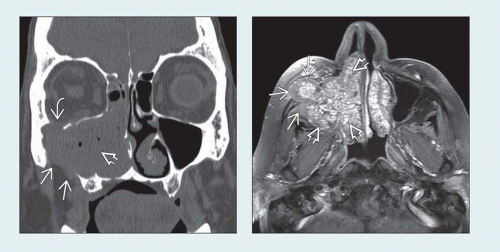
 (Left) Coronal bone CT shows a mass
 filling the right nasal cavity and maxillary sinus with erosion of the orbital floor filling the right nasal cavity and maxillary sinus with erosion of the orbital floor  and lateral wall of the maxillary sinus and lateral wall of the maxillary sinus  . (Right) Axial T1WI C+ FS MR in the same patient shows typical cerebriform appearance of inverted papilloma throughout most of the tumor mass . (Right) Axial T1WI C+ FS MR in the same patient shows typical cerebriform appearance of inverted papilloma throughout most of the tumor mass  . Note loss of this appearance at more solid portion of mass at anterolateral margin . Note loss of this appearance at more solid portion of mass at anterolateral margin  , which invades through to cheek. This correlated with areas of carcinoma found at surgical resection. , which invades through to cheek. This correlated with areas of carcinoma found at surgical resection.Stay updated, free articles. Join our Telegram channel
Full access? Get Clinical Tree
 Get Clinical Tree app for offline access

|