Hereditary cancer syndromes are characterized by increased susceptibility to early-onset development of multifocal, organ-specific, and pathogenetically unique tumors. Commonly inherited as autosomal dominant disorders, these syndromes are associated with germline mutations of single genes that function as tumor suppressor genes. Recent advances in genetics and pathology have thrown fresh light on the molecular biology, tumor pathways, and behavior of distinctive tumors that occur in these syndromes. Comprehensive investigations of these rare syndromes have led to a better understanding of the pathogenesis and management of the more common sporadic neoplasms, including deciphering of driver mutations, tumor pathways, natural history, and prognosis.
Key points
-
•
Patients with a diverse spectrum of germline mutation-driven, multisystem, hereditary cancer syndromes demonstrate predisposition to early-onset development of multifocal, phenotypically distinctive, benign and malignant tumors.
-
•
Accurate diagnosis depends on a constellation of clinical symptoms, laboratory abnormalities, cross-sectional imaging findings, histopathology, and genetic tests.
-
•
Management is complicated due to occurrence of multiple, deeply located tumors, and clinical presentation with locally advanced or metastatic disease.
-
•
Early diagnosis and optimal management of patients with inherited tumors are dependent on several factors: family history, genetic tests, and prompt institution of clinical/laboratory/imaging-based screening paradigms.
-
•
Improved understanding of hereditary syndromes has helped elucidate the pivotal role of genes in tumor pathogenesis, thus paving the way for better molecular diagnostics and pathway-specific targeted therapeutics.
Abbreviations
| ccRCCs | clear cell renal cell carcinomas |
| CHRPE | congenital hypertrophy of retinal pigment epithelium |
| CMP | corticomedullary phase |
| CRCs | colorectal cancers |
| DF | desmoid fibromatosis |
| EGFR | epidermal growth factor receptor |
| FAP | familial adenomatous polyposis |
| FH | fumarate hydratase |
| HGSCs | high-grade serous carcinomas |
| HIFs | hypoxia-inducible factors |
| HLRCC | hereditary leiomyomatosis and renal cell carcinoma |
| HNPCC | hereditary nonpolyposis colorectal cancer |
| LS | Lynch syndrome |
| MMR | mismatch repair |
| pVHL | VHL protein |
| RCC | renal cell carcinoma |
| VEGF | vascular endothelial growth factor |
| VHL | von Hippel-Lindau syndrome |
Introduction
A heterogeneous group of hereditary syndromes is characterized by a predisposition to a multitude of pathologically exclusive neoplasms that display specific organ distribution and show familial aggregation. Benign and malignant hereditary tumors comprise 3% to 10% of neoplasia in several visceral organs. These inherited tumors exhibit an earlier age of onset than sporadic tumors and affect a multitude of organs. ,, As a representative example, patients with von Hippel-Lindau syndrome (VHL) demonstrate increased proclivity for the development of central nervous system/retinal hemangioblastomas, pheochromocytomas, clear cell renal cell carcinomas (ccRCCs), and pancreatic neuroendocrine tumors. Hereditary syndromes are commonly inherited in an autosomal dominant pattern and result from single-gene mutations of tumor suppressor genes. Biallelic gene inactivation, according to classic Knudson’s 2-hit model, results in tumorigenesis. The diagnosis of these syndromes is based on family history, discovery of distinctive neoplasms with characteristic histopathology, laboratory abnormalities, imaging features, and genetic tests. A wide array of cross-sectional imaging modalities plays a pivotal role in the detection, characterization, and localization of specific tumors, as well as allowing robust surveillance strategies following treatment. Although imaging characteristics are pathognomonic for some neoplasms, histopathological examination is required for confirmation of the diagnosis. A select group of recently described molecularly defined tumors warrants advanced genetic testing to establish the diagnosis. Imaging is also an integral component of standardized screening protocols employed for early diagnosis, thereby facilitating optimal management while improving prognosis. Patients with hereditary tumor syndromes benefit from evaluation by a multidisciplinary team in specialized centers.
Comprehensive investigation of hereditary cancer syndromes have shed light and provided unique insights into the role of genetic/epigenetic mutations in molecular tumorigenesis, tumor pathways, and the biological behavior of the more common sporadic neoplasms. The identification and mapping of the tumor suppressor VHL gene led to novel insights into the pivotal role of VHL protein (pVHL) in oxygen sensing as well as regulation of hypoxia-inducible factors (HIFs) and downstream angiogenic/metabolic/somatic growth pathways. Based on the central role of the pVHL-HIF pathway in the pathogenesis of ccRCCs, a plethora of novel therapeutic agents targeting the myriad tumor pathways are currently being effectively used in patients with advanced and metastatic ccRCCs. Seminal studies on the tubal origin of most BRCA-associated high-grade serous carcinomas (HGSCs) led to similar conclusions for many sporadic HGSCs as well. , As a result, prophylactic salpingo-oophorectomy and opportunistic salpingectomy are being offered to reduce ovarian cancer risk in both hereditary and sporadic scenarios. , Increasing use of poly-adenosine diphosphate (ADP)-ribose polymerase inhibitors based on BRCAness of ovarian cancers and synthetic lethality is another example wherein knowledge gleaned from rare hereditary syndromes has beneficially impacted innumerable patients with sporadic cancers.
Hereditary syndromes can be classified based on the predominant involvement of organ systems or specific tumor phenotypes, such as multiple endocrine neoplasia and hereditary pheochromocytoma/paraganglioma syndromes. As the topic is extensive, we limit our article to syndromes that primarily involve the kidneys, colon, and ovaries. Additionally, within each category, we will discuss only selected syndromes, as comprehensive coverage of all syndromes is beyond the scale and scope of this article.
Hereditary renal tumor syndromes
There is a diverse spectrum of hereditary syndromes that predispose patients to renal tumors of characteristic cytogenetics, histomorphology, cross-sectional imaging findings, and biological behavior. ,, Patients with these syndromes commonly develop bilateral, multifocal renal tumors, except for hereditary leiomyomatosis and renal cell carcinoma (HLRCC) syndrome, which typically shows solitary tumors. Barring hereditary papillary RCC syndrome, which is associated with the promotion of the c-MET proto-oncogene, these syndromes are characterized by the silencing of the tumor suppressor genes. Apart from Birt-Hogg-Dube syndrome, which commonly shows a broad spectrum of renal tumors, the inherited syndromes are characterized by the development of one specific histopathology of renal tumors. A synopsis of hereditary renal tumor syndromes is presented in Table 1 . Germline testing is recommended in a variety of clinical scenarios that include a diagnosis of a renal tumor in the context of a family history of hereditary syndromes, a diagnosis of RCC in any patient younger than 45 years, bilateral or multiple renal masses, and a spectrum of characteristic skin or central nervous system (CNS) tumors. Screening protocols for other tumors of multisystem disorders differ from screening suggestions/recommendations for renal tumors, which are summarized in Table 2 . MR imaging is the preferred modality for screening individuals at risk for hereditary syndromes; ultrasonography and CT may be considered on an individual basis.
Table 1
Hereditary renal tumor syndromes
| Syndrome | Gene (Location) | Protein Functions | Renal Tumor | Extrarenal Manifestations |
|---|---|---|---|---|
| VHL | VHL (3p25.3) | Ubiquitination and proteasomal degradation of proteins, including HIFs. | ccRCCs | CNS/retinal hemangioblastomas, pheochromocytomas, pancreatic neuroendocrine tumors, and endolymphatic sac tumors |
| Hereditary papillary RCC | MET proto-oncogene, a member of the receptor tyrosine kinase family (7q31) | After binding with hepatocyte growth factor, c-MET promotes cellular survival, migration, and invasion. | Papillary RCCs | |
| HLRCC | FH (1q42.3-q43) | FH is a key mitochondrial enzyme associated with Kreb’s cycle. FH-deficiency leads to accumulation of fumarate and other oncometabolites. | FH-deficient RCCs | Skin and uterine leiomyomas |
| Birt-Hogg-Dube | Folliculin (17q11.2) | Folliculin loss may activate multiple cellular signaling pathways, including mTOR pathway. | Hybrid oncocytic tumors, oncocytomas, and chromophobe RCCs | Skin fibrofolliculomas, lung cysts predisposing to pneumothorax |
| SDH-deficient tumor syndromes | SDHA, SDHB, SDHC, SDHD, SDHAF2 | The SDH complex is a critical mitochondrial enzyme linked with the Krebs’ cycle and the electron transport chain. Accumulation of succinate, an oncometabolite involved in diverse tumor pathways. | SDH-deficient RCCs | Pheochromocytomas, paragangliomas, and gastric gastrointestinal stromal tumor |
| Tuberous sclerosis | TSC1, TSC2 (9q34, 16p13.3) | Hamartin and tuberin are encoded by TSC1 and TSC2. Gene mutations result in upregulation of mTOR signaling, leading to tumorigenesis. | Angiomyolipomas, RCCs, cysts |
CNS tubers, hamartomas, and subependymal giant cell astrocytomas.
Cardiac rhabdomyomas, skin nodules, facial angiofibromas, and lymphangioleiomyomatosis |
| BAP1 tumor predisposition syndrome | BAP1 (3p21.1) | BAP1 protein interacts with many proteins that are involved in DNA damage response, cell cycle regulation, and metabolism. | RCC | Melanocytic tumors, melanoma, mesotheliomas, and basal cell carcinoma |
Abbreviation: SDH, succinate dehydrogenase.
Table 2
Screening suggestions/recommendations for hereditary renal tumors
| Syndrome | Suggested Age to Start Screening (Years) | Suggested Imaging Intervals |
|---|---|---|
| VHL | 15 | Every 2 y |
| Hereditary papillary RCC | 30 | Every 1–2 y |
| HLRCC | 8–10 | Every year |
| Birt-Hogg-Dube | 20 | Every 3 y |
| SDH-deficient tumor syndromes | 12 | Every 4–6 y |
| Tuberous sclerosis | 12 | Every 3–5 y |
| BAP1 tumor predisposition syndrome | 20 | Every 2 y |
von Hippel-Lindau syndrome
VHL syndrome is an autosomal dominant disorder (with a penetrance of >90%) caused by inactivating germline mutations of the VHL tumor suppressor gene, located on chromosome 3p25.3. Approximately 80% of patients have an affected parent. VHL is characterized by the development of visceral cysts and cystic neoplasms, as well as a broad spectrum of distinctive benign and malignant neoplasms by the age of 30 years ( Fig. 1 ). VHL can be broadly categorized into 2 clinical subtypes. While type I disease, caused by large deletions or truncation mutations, is associated with increased predisposition for retinal and craniospinal axis hemangioblastomas and RCCs, type II disease is associated with missense mutations and increased proclivity for pheochromocytoma. The genitourinary tract manifestations of VHL include bilateral, multiple renal cysts of varying complexity (70% of patients), ccRCCs, and epididymal cystadenomas. Bilateral epididymal cystadenomas are virtually pathognomonic of the syndrome.
( A , B ) Coronal, nonfat-saturated, T2-weighted MR image of the kidneys ( A ) in a 25-year-old man with VHL shows bilateral renal cell carcinomas ( blue arrows ). Scattered, small, bilateral renal cysts ( red arrows ) are also seen. Cerebellar hemangioblastomas in the same patient with VHL. Sagittal, postcontrast T1-T1-weighted MR image ( B ) shows a cerebellar cyst ( blue arrow ) that exhibits a hypervascular mural nodule ( red arrow ). An adjacent avidly enhancing solid hemangioblastoma ( green arrow ) is also seen.
About 70% of VHL patients show a propensity for early onset development of bilateral and multicentric ccRCCs. The ccRCC is the prototype histopathological subtype of RCCs in patients with VHL; the average patient age is 40 to 45 years, about a decade earlier than patients with sporadic tumors. At histopathology, ccRCC is comprised of nests of tumor cells with optically clear cytoplasm that is rich in glycogen and lipid; the distinctive lipid deposition is due to the HIF-dependent downregulation of carnitine palmitoyltransferase 1A. A profuse network of thin-walled, sinusoid-like capillaries is characteristic and is presumably driven by overexpression of vascular endothelial growth factor (VEGF)-A. The ccRCCs may manifest as complex cystic masses with enhancing mural nodules or heterogeneously enhancing, hypervascular, and expansile solid masses. Solid ccRCCs typically demonstrate avid contrast enhancement on corticomedullary phase (CMP) images with de-enhancement on delayed phase images. , On MR imaging, ccRCCs show heterogeneously hyperintense on T2WI and show hypervascularity on CMP images , ( Fig. 2 ). Subtraction techniques enable the assessment of the degree of enhancement, particularly in T1-hyperintense lesions, as well as a more accurate evaluation of residual/recurrent disease following surgery or ablation. Although lipid-rich tumor cells are commonly seen at histopathology, the detection of microscopic fat in ccRCCs on MR imaging is unusual. Quantitative signal loss on chemical shift MR imaging demonstrates high-specificity and accuracy for detecting microscopic fat in ccRCCs. The ccRCCs show marked propensity for intravascular growth into the renal vein, inferior vena cava, and the right atrium. Also, ccRCCs are biologically aggressive with frequent metastases to the lung, liver, lymph nodes, bones, and pleura in descending order of frequency. ,
( A – C ) Axial, fat-saturated T2-weighted MR image ( A ) in a 26-year-old man with VHL-associated ccRCC shows a well-circumscribed hyperintense mass in the left kidney ( blue arrow ). Axial, postcontrast, fat-saturated, T1-weighted MR image during corticomedullary phase ( B ) displays hypervascularity of the soft tissue mass. There is heterogeneous de-enhancement of the mass ( blue arrow ) on the nephrographic phase image ( C ).
Metastatic ccRCC is the leading cause of death in patients with VHL, accounting for 35% to 45% of deaths. Until recently, clinical management of VHL-associated RCCs consisted of imaging surveillance of tumors till one of the tumors met the threshold size of 3 cm for surgical resection or ablation , ( Fig. 3 ). Belzutifan, a small-molecule HIF-2α inhibitor, is the first and, to date, the only US Food and Drug Administration (FDA)-approved drug for prophylactic systemic therapy in VHL patients. It is well-tolerated with a long-term safety profile and durable antitumor activity and efficacy in shrinking multisystem tumors, including RCCs, resulting in a lower need for multiple surgical interventions , ( Fig. 4 ). Sixty-seven percent of study participants with VHL-associated RCCs in a recent study showed an objective response of at least 42 months duration (70% of cases) with a favorable safety profile.
( A , B ) A 44-year-old woman with VHL-associated ccRCC. Axial, fat-suppressed, postcontrast T1-weighted MR image ( A ) demonstrates a heterogeneously enhancing right kidney expansile solid mass ( blue arrow ). Postablation changes ( blue arrow ) within the right kidney on axial contrast-enhanced T1-weighted MR image ( B ) subtraction images (not shown) did not show nodular enhancement to suggest recurrence.
( A , B ) A 55-year-old woman with VHL-associated ccRCC. Axial contrast-enhanced T1-weighted MR image ( A ) depicts an enhancing, left kidney mass ( blue arrow ). Axial contrast-enhanced T1-weighted MR image ( B ) 2 years after belzutifan therapy shows an interval decrease in the size of the reference mass ( blue arrow ).
Hereditary leiomyomatosis and renal cell carcinoma syndrome
Also referred to as Reed syndrome, HLRCC syndrome is an autosomal dominant tumor predisposition syndrome, caused by heterozygous germline mutations involving the fumarate hydratase (FH) gene located on 1q42.3-q43. , The syndrome is characterized by the development of cutaneous leiomyomas (up to 75% of patients), multiple uterine leiomyomas (up to 80% of patients), and FH-deficient RCCs (15%–30% of patients). Leiomyomas occur 10 to 15 years earlier than RCCs. The median age of onset for RCC is 44 years.
The FH gene encodes the FH protein, a key enzyme in the mitochondrial tricarboxylic acid (Krebs) cycle, which generates ATP through oxidative phosphorylation. The metabolic and oncogenic consequences of FH deficiency include pseudohypoxia, promotion of aerobic glycolysis and glutamine catabolism (Warburg effect), constitutive stabilization of HIFs, protein succination, as well as impaired homologous recombination-dependent DNA damage repair. ,, HIF-related downstream pathways with increased transcription of VEGF as well as epidermal growth factor receptor (EGFR) signaling, play significant roles in tumorigenesis. FH-deficient neoplasms exhibit a distinctive histomorphology characterized by pleomorphic, admixed architectural patterns, with the papillary configuration being the most common. The cells show abundant eosinophilic cytoplasm and a large, solitary, inclusion-like macronucleoli with perinucleolar halos reminiscent of cytomegalovirus (CMV) inclusion bodies. Identification of the FH mutation by genetic tests or immunohistochemistry (IHC) clinches the diagnosis. ,
FH-deficient RCCs are currently classified under the rubric of molecularly defined RCCs according to the 2022 World Health Organization (WHO) classification schema for renal tumors. , FH-deficient RCC typically manifests as a large, unilateral, solitary, solid or solid and cystic tumor with a distinct male preponderance (M: F = 1.9:1) ( Fig. 5 ). Characteristic imaging features include hypovascularity and marked restricted diffusion. They are biologically aggressive with frequent regional metastatic lymphadenopathy and perinephric metastases in 50% of cases and warrant prompt radical nephrectomy even for small tumors. , Significant risk for metastatic disease and predisposition to RCC development in the other kidney exists even after nephrectomy. Clinical management of patients consists of imaging surveillance to detect renal tumors when they are small so that prompt surgery may be instituted. Eighty percent of patients may demonstrate metastases either at presentation or within 3 years of diagnosis. FH-deficient RCCs are refractory to standard chemotherapy; immune checkpoint inhibitors, anti-VEGF, and anti-EGFR drugs are being used to treat these patients. A recent study found that a combination of bevacizumab and erlotinib resulted in 72% response rates in patients with HLRCC-associated RCCs. Despite encouraging results, the diagnosis portends a poor prognosis with dismal survival rates.
( A – C ) A 46-year-old woman with HLRCC syndrome. Coronal, nonfat suppressed, T2-weighted MR image of the kidneys ( A ) shows multiple uterine leiomyomas ( blue arrows ) and a left kidney hypointense mass ( red arrow ) with associated left renal vein tumor thrombus ( green arrow ). Axial, fat-suppressed, T2-weighted MR image ( B ) depicts an infiltrative left kidney malignancy ( red arrow ) with an expansile thrombus within the left renal vein ( green arrow ). Axial contrast-enhanced CT of the abdomen ( C ) demonstrates biopsy-proven fumarate hydratase-deficient left kidney renal cell carcinoma ( red arrow ) with associated tumor thrombus ( green arrow ).
Hereditary colorectal cancer syndromes
Approximately 10% of colorectal cancers (CRCs) occur in the context of a broad spectrum of polyposis and nonpolyposis syndromes caused by germline mutations of a plethora of genes. The polyposis syndromes characterized by diverse genetic abnormalities include adenomatous (classic/attenuated FAP, and non-FAP syndromes), hamartomatous (juvenile polyposis, Peutz-Jeghers, Cowden, and PTEN hamartoma), serrated (serrated polyposis syndrome, RNF43 -associated polyposis), and mixed polyposis syndromes (BMPR1A-associated polyposis). A synopsis of the 2 most common hereditary CRC syndromes is presented in Table 3 .
Table 3
Common hereditary colorectal cancer syndromes
| Syndrome | Gene | Protein Functions | Colon Cancer | Extracolonic Manifestations |
|---|---|---|---|---|
| LS |
MLH1
(3p21)
MSH2 (2p16) MSH6 (2p16) PMS2 (7p22) EPCAM |
DNA proofreading, excision, and repair of mismatch errors during DNA replication | Colorectal cancer |
Endometrial cancer, Ovarian cancer, and small bowel and gastric cancer
Urothelial cancer and pancreatic cancer |
| FAP | APC | Degrades β-catenin oncoprotein in the absence of a Wnt ligand | Colorectal cancer | Duodenal cancer, congenital nodular thyroid gland, desmoid fibromatosis, osteomas, papillary thyroid carcinoma, hepatoblastoma, brain tumors, dental abnormalities, lipoma, adrenal cortical carcinomas, and nasopharyngeal angiofibroma |
Lynch syndrome
Previously known as hereditary nonpolyposis colorectal cancer (HNPCC), Lynch syndrome (LS) is the most frequent CRC syndrome, with a lifetime risk of colon cancer up to 80% and accounts for 2% to 4% of all CRC cases. LS is an autosomal dominant disorder caused by a mutation in one of the DNA mismatch repair (MMR) genes, most commonly MLH1 , MSH2 , MSH6 , and PMS2. , Deletion of the EPCAM gene, accompanied by silencing or epigenetic changes of the MSH2, is another mutation, albeit less frequent. , MMR genes encode proteins that are responsible for DNA proofreading, excision, and repair of mismatch errors during DNA replication. Loss-of-function mutations in LS lead to the hallmark feature of microsatellite instability and subsequent development of cancers characterized by defective DNA repair of replication errors. , Different LS mutations are characterized by variable risk profiles for a broad spectrum of multiorgan malignancies. Approximately 70% to 85% of LS cases are due to mutations in MLH1 and MSH2, which carry the highest risk. The diagnosis of LS can be achieved either by polymerase chain reaction amplification of microsatellite repeats or by next-generation sequencing. ,
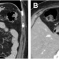
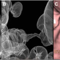
LS-associated CRCs occur in patients with a mean age of 44 to 61 years and exhibit a distinct predilection for the right colon in 60% to 80% of cases ( Fig. 6 ). Imaging plays a crucial role in the diagnosis, treatment, assessment, and surveillance of LS ( Fig. 7 ). CRC manifests as circumferential wall thickening or an intraluminal polypoid mass on computed tomography (CT) or MR imaging. Contrast-enhanced CT study is crucial for locoregional staging as well as assessment of visceral metastases; flourodeoxyglucose (FDG) PET/CT is superior in detecting overall tumor burden. FDG PET/CT is also helpful in evaluating treatment response and for posttreatment surveillance. LS-associated CRCs have a better prognosis than sporadic cases due to active host immune defense mechanisms and decreased tumor cell survival in microsatellite instability (MSI) tumors.