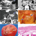
Fig. 6.1
Serous cystadenomas macroscopic appearance. Photographs of four resected gross specimens (a–d) show the characteristic lobulated smooth surface of these multicystic masses
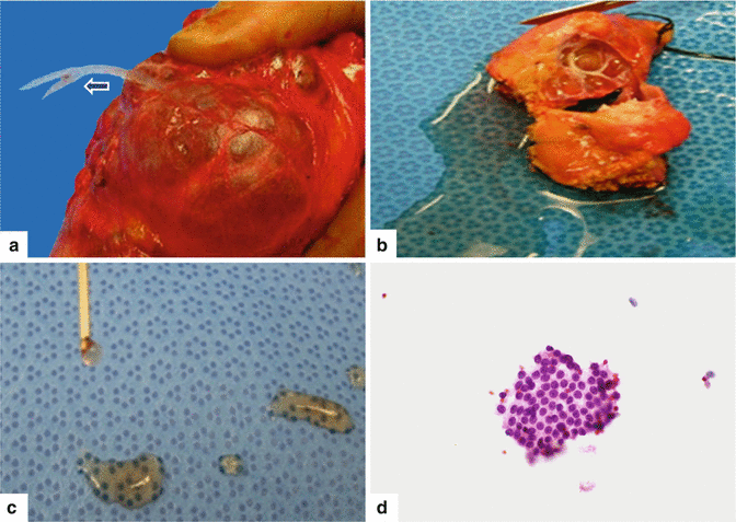
Fig. 6.2
Fluid characteristics in serous cystadenoma. These multiloculated cystic masses are filled with watery serous fluid that can be clear (arrow) to brown. The photographs depicted in this figure (a, b) demonstrate the characteristic thin, non-viscous, transparent fluid (c), which may contain (d) floating clusters of benign-appearing epithelial tumor cells (H&E, 60×)
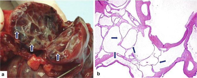
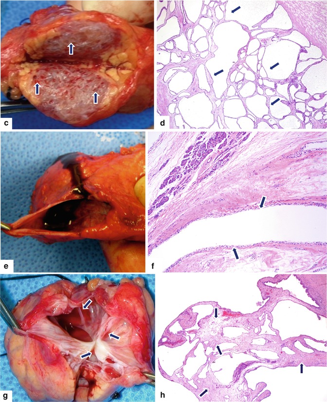
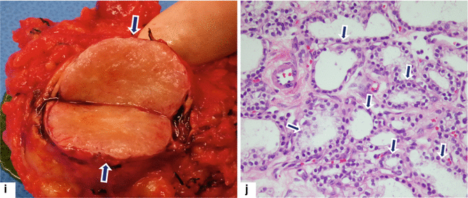
Fig. 6.3
Serous cystadenomas gross internal appearance. Gross and microscopic photographs of four different bivalved serous cystadenomas show (a, b) (H&E, 4×) a mass composed by grape-like clusters of cysts with a fibrous septa and smooth inner lining (arrows).(c, d) (H&E, 4×) a mass composed of multiple minute cysts arranged in a honeycomb fashion (arrows) and (e, f) (H&E, 10×) a single unilocular cyst with smooth inner surface (arrows) filled with dark brown watery fluid. (g, h) (H&E, 4×) an oligocyst with few internal septations (arrows). (i, j) (H&E, 4×) a mass with fleshy appearance (arrows) composed of multiple, minute cysts (arrows)
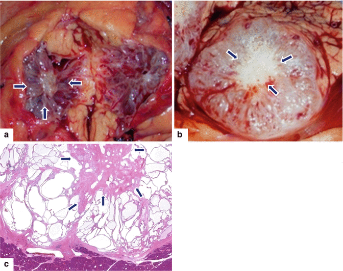
Fig. 6.4
Central scar in serous cystadenomas. Photographs of two bivalved serous cystadenomas (a, b) demonstrate the characteristic fibrous central scar, radiologically known as the “sunburst” sign (arrows). Microscopically, the central scar is composed of dense fibroconnective tissue (c) (arrows) (H&E, 4×)
Variable size, round to ovoid, sharply outlined cystic mass that can have a lobulated, irregular, or smooth serosal surface
Microcystic: Grape-like or sponge-like cluster of thin-walled cysts with fibrous septa or central scar (sunburst appearance)
Oligocystic: Single cystic mass, with or without internal septations
Filled with clear, watery, straw-colored fluid
6.3.2 Microscopic Appearance
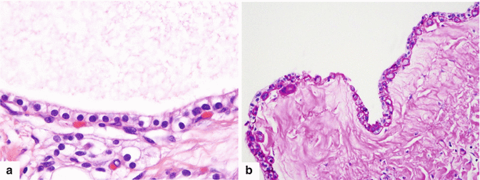
Fig. 6.5
Lining epithelium in serous cystadenomas. The serous epithelium lining the cysts is composed of small cuboidal cells with clear cytoplasm (a) (H&E, 60×) rich in glycogen (b) (PAS stain, 60×), and round central hyperchromatic nuclei
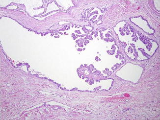
Fig. 6.6
Serous epithelium with small papillary projections (H&E, 10×)
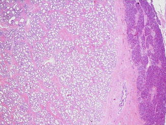
Fig. 6.7
Serous cystadenomas can have a more solid pattern when the tumor is composed predominantly by minute microcysts (H&E, 10×)
Cysts lined by flat, cuboidal epithelium composed of small cells with round to oval nuclei and clear to pale, well-defined cytoplasm rich in glycogen
Small papillary projections can be present.
Rich capillary network intimately admixed with epithelium.
A myoepithelial cell layer can be observed between the epithelium and the collagenous stroma.
6.4 Clinical Presentation
Most patients are asymptomatic (found incidentally by imaging)
Vague abdominal pain
Malaise
Rarely jaundice
Palpable mass
6.5 Laboratory Evaluation
Typically nonspecific
Serum markers:
CA 19–9 and CEA normal
Aspirated fluid:
Low CEA < 5 ng/ml, amylase negative
Practical Pearls
An elevated CEA may be detected from the aspirated SCA fluid in rare instances (false positive).
The presence of an elevated amylase in aspirated fluid from a unilocular cystic pancreatic mass excludes the possibility of an oligocystic serous cystadenoma.
6.6 Imaging
Preferred imaging modalities
Multidetector computed tomography (MDCT)
Magnetic resonance imaging (MRI) with MR cholangiopancreatography (MRCP)
Endoscopic ultrasound (EUS)
SCA imaging patterns:
Polycystic
Honeycombing
Oligocystic (unilocular)
Practical Pearls
Although the mean size of SCA is ± 5 cm, these cystic tumors may reach >25 cm.
Synchronous pancreatic masses may be found associated with SCA.
Multiple SCAs of different sizes may be found sporadically or associated with von Hippel-Lindau syndrome.
6.6.1 Ultrasound (US) (Figs. 6.8–6.19)
Transabdominal ultrasound, endoscopic ultrasound (EUS), and intraoperative ultrasound (IOUS)
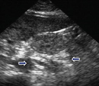
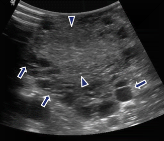

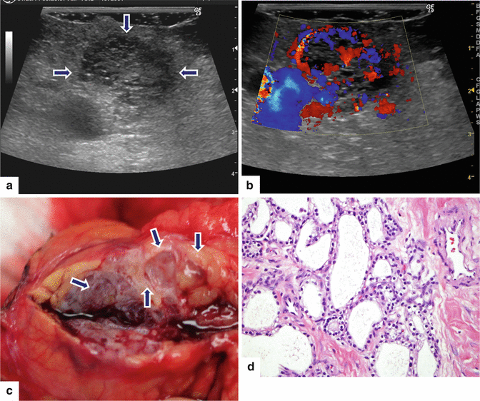
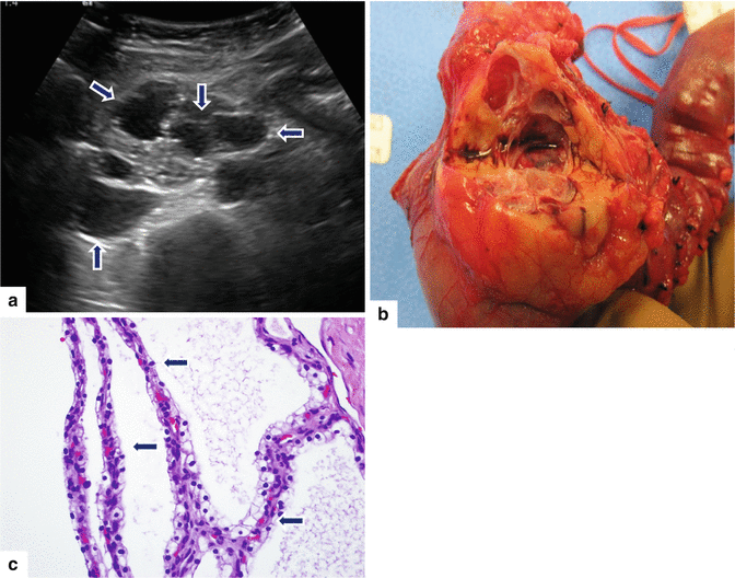
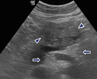
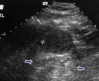
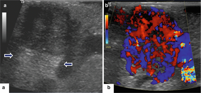
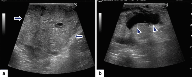
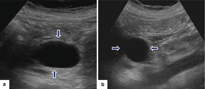
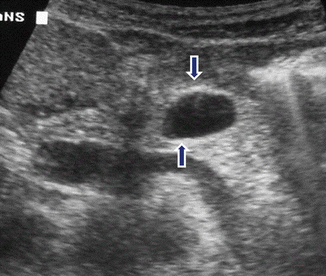
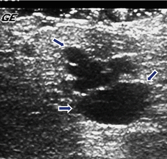
Fig. 6.8
Serous cystadenoma (microcystic type) on transabdominal ultrasound. Incidental finding in a 67-year-old woman with mild abdominal discomfort. Transverse image of the head of the pancreas shows a lobulated echogenic mass in the head of the pancreas composed of small cysts and increased posterior enhancement of the sound (arrows)
Fig. 6.9
Serous cystadenoma (microcystic type) on intraoperative ultrasound (IOUS). A 48-year-old female patient with epigastric pain. Transverse image demonstrates a round, lobulated mass with multiple, peripherally small cysts (arrows) and an echogenic central scar (arrowheads)
Fig. 6.10
Serous cystadenoma (microcystic type) on intraoperative ultrasound (IOUS). A 65-year-old female with a known serous cystadenoma stable for 25 years. The patient underwent an intraoperative ultrasound of the liver for the evaluation of liver metastases from colon adenocarcinoma and, at the same time, the pancreatic mass was evaluated with IOUS. Transverse image (a) and color Doppler ultrasound (b, c) show a round hyperechoic mass with small peripheral small cysts (arrows) and increased posterior enhancement of the sound involving the body of the pancreas (arrows). Note the prominent arterial flow in the central scar demonstrated by color Doppler ultrasound (b, c). This size of the lesion was compared with a non-contrast-enhanced CT performed 3 years before (d). No change in the size of this pancreatic cystic mass was noted (arrows)
Fig. 6.11
Serous cystadenoma (microcystic type) on transabdominal ultrasound. A 55-year-old female with left upper quadrant discomfort. Transverse image (a) shows a complex mass in the body of the pancreas, composed of multiple, small cysts, separated by an echogenic, fibrous scar (arrows) and with increased posterior acoustic enhancement. Color Doppler ultrasound (b) shows the presence of multiple small arteries within the central scar. Patient underwent a distal pancreatectomy. Photograph of the gross specimen (c) shows a cystic mass composed of multiple small cysts (arrows). Histological section (d) shows small cysts lined by clear, cuboidal epithelium (H&E, 4×)
Fig. 6.12
Serous cystadenoma (microcystic type) on transabdominal ultrasound. A 38-year-old female with epigastric pain found to have a cystic lesion in the head of the pancreas. Endoscopic ultrasound with fine-needle aspiration suggested the possibility of a mucinous cystic neoplasm. Transverse image (a) demonstrates a pancreatic mass with multiple small cysts of different sizes involving the head of the pancreas (arrows). The patient underwent a Whipple procedure. Photograph of the gross specimen (b) reveals a well-circumscribed, round mass showing small cysts with a smooth lining. Microscopic examination of the mass (c) (H&E, 10×) reveals that the lining was composed of small cuboidal cells with clear cytoplasm and round nuclei (arrows), representing a serous cystadenoma, microcystic type
Fig. 6.13
Serous cystadenoma (honeycomb type) on transabdominal ultrasound. Incidental finding in a 41-year-old female patient evaluated with ultrasound for possible gallstones. Transverse image shows a round heterogeneous hyperechoic mass involving the body of the pancreas (arrowheads). Note the posterior acoustic enhancement (arrows)
Fig. 6.14
Serous cystadenoma (honeycomb type) on transabdominal ultrasound. A 53-year-old male with mild epigastric pain. Transverse image shows a homogeneous, hypoechoic, ovoid mass (stars) with posterior acoustic enhancement (arrows) involving the body of the pancreas
Fig. 6.15
Serous cystadenoma (honeycomb type) on intraoperative ultrasound. A 49-year-old female with epigastric pain and weight loss. Serum markers were negative. Transverse image (a) demonstrates a round, heterogeneous mass with posterior acoustic enhancement involving the head of the pancreas (arrows). Color Doppler ultrasound (b) demonstrates high vascularity within this mass
Fig. 6.16
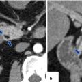
Serous cystadenoma (honeycomb type) associated with pancreatic duct dilatation on intraoperative ultrasound (IOUS). An 81-year-old female patient in whom an incidental pancreatic mass was discovered on a CT of the abdomen performed to evaluate abdominal trauma. IOUS transverse scans show a heterogeneous, hyperechoic, lobulated mass (arrows) involving the neck of the pancreas (a). Note the dilatation of the pancreatic duct distal to this mass (b) (arrowheads). The patient underwent a distal pancreatectomy and splenectomy. Final pathology: serous cystadenoma associated with chronic pancreatitis involving the distal pancreas
Fig. 6.17
Serous cystadenoma (oligocystic type) on intraoperative ultrasound (IOUS). Incidental finding in a 45-year-old female with right upper quadrant pain. Transverse (a) and sagittal (b) images show an ovoid cystic mass in the neck of the pancreas (arrows)
Fig. 6.18
Serous cystadenoma (oligocystic type) on intraoperative ultrasound (IOUS). Incidental finding in a 45-year-old female, who subsequently had an endoscopic ultrasound with fine-needle aspiration which was non-diagnostic. Serum markers were negative. Patient underwent a central pancreatectomy. Transverse image demonstrates an ovoid cystic mass involving the body of the pancreas (arrows)
Fig. 6.19
Serous cystadenomas (oligocystic type) on intraoperative ultrasound (IOUS). A 48-year-old female presenting with vague epigastric pain. Transverse image demonstrates a lobulated, septated cystic mass involving the neck of the pancreas (arrows)
Microcystic/honeycombing types:
Lobulated cystic mass composed by a cluster like grape cysts
Round or ovoid hyperechoic or hypoechoic, lobulated mass with increased posterior enhancement
Additional findings:
Central stellate-shaped echogenic area (central scar) better identified on intraoperative or endoscopic ultrasound
Amorphous, central, echogenic foci (calcifications)
On color Doppler: hypervascular septa and capsule
Oligocystic type:
Round or ovoid, unilocular cystic mass, with thin walls, and with or without internal septations
Practical Pearls
The microcystic/honeycombing type of serous cystadenoma may be confused with a solid mass by ultrasound due to the numerous subcentimeter cysts not being able to be individually distinguished by this imaging modality.
The key to establishing this diagnosis is the detection of posterior acoustic enhancement associated with a well-defined, loculated, echogenic, pancreatic mass.
6.6.2 Computed Tomography (CT) (Figs. 6.20–6.48)
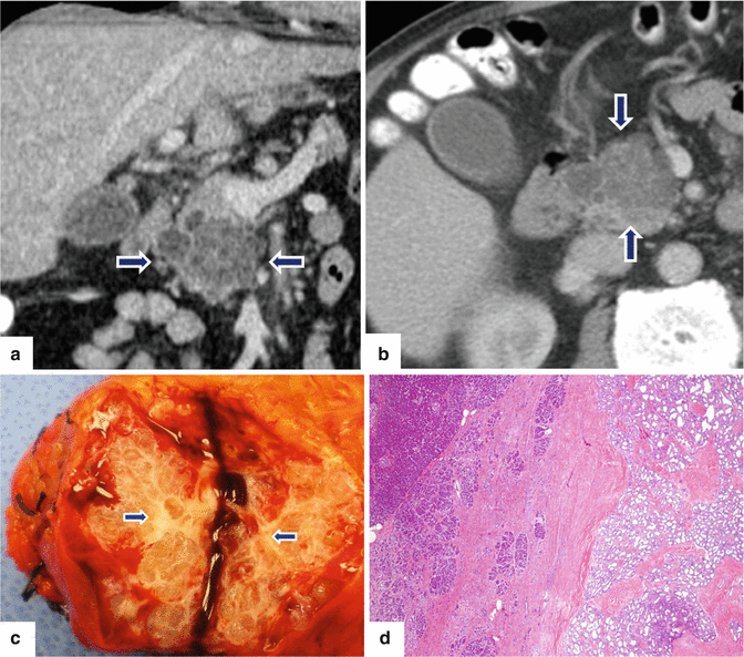
Fig. 6.20
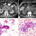
Serous cystadenoma (microcystic type) on CT. An 82-year-old male with abnormal liver function tests and an incidental finding on screening abdominal CT. CECT coronal (a) and axial (b) images show a lobulated mass involving the head of the pancreas composed of multiple, small cysts of different sizes divided by fibrous septa (arrows). Photograph of the sectioned gross specimen (c) demonstrates a well-defined, fairly round, firm mass composed of multiple small cysts. A small, white, fibrotic central scar (arrows). Histological section (d) (H&E, 4×) shows a serous cystadenoma separated from adjacent pancreatic parenchyma by fibrous tissue
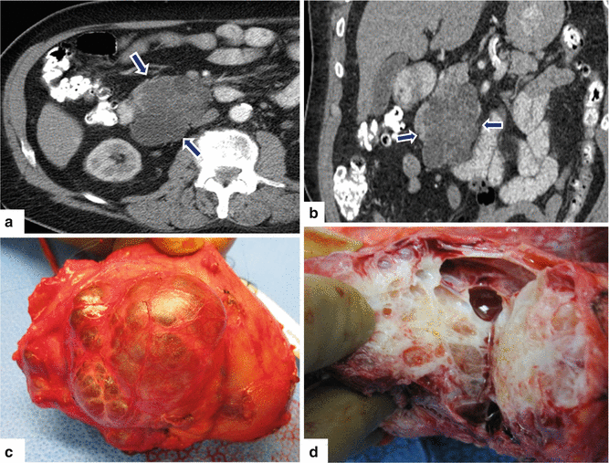
Fig. 6.21
Serous cystadenoma (microcystic type) on CT. A 72-year-old male patient with abdominal discomfort. CECT axial (a) and coronal (b) images demonstrate an ovoid cystic mass (arrows) with internal, thin septa in the head of the pancreas. Photographs of the gross specimen (c) demonstrate a lobulated cystic mass. Upon dissection (d), the multicystic mass with thick fibrotic septa and clear serous fluid was identified
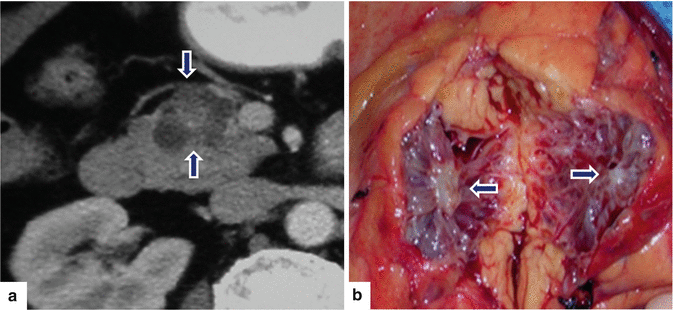
Fig. 6.22
Serous cystadenoma (microcystic type) on CT. Incidental finding in a 68-year-old female patient. CECT axial image (a) shows a small, lobulated, multicystic mass with a central scar involving the head of the pancreas (arrows). Photograph of the bivalved gross specimen (b) shows an irregular cystic mass with a prominent, tan-white, stellate-like. Central scar (arrows)
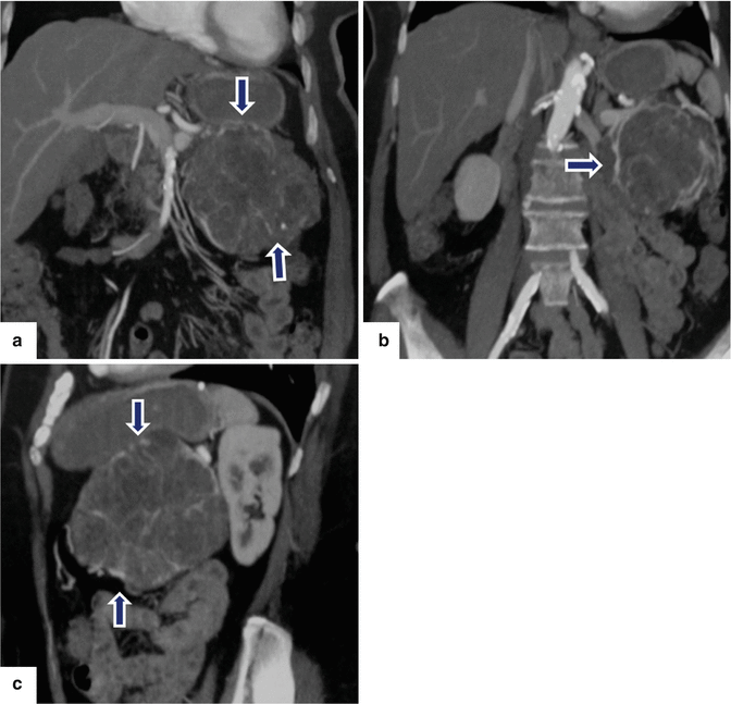
Fig. 6.23
Serous cystadenoma (microcystic type) on CT. An 80-year-old female patient with mild epigastric pain. CECT coronal (a, b) and sagittal (c) images show a large, lobulated multicystic mass with an enhancing central fibrous scar (arrows)
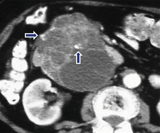
Fig. 6.24
Serous cystadenoma (microcystic type) with coarse calcifications on CT. A 67-year-old female patient with abdominal discomfort. CECT axial image shows a lobulated multicystic mass in the head of the pancreas with central and peripheral coarse calcifications in (arrows)
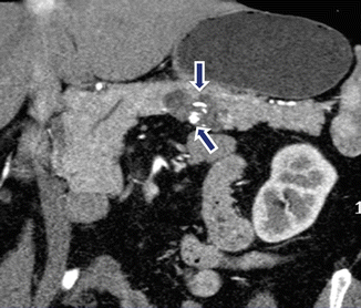
Fig. 6.25
Serous cystadenoma (microcystic type) with coarse calcifications on CT. Incidental finding on a 71-year-old female. CECT coronal image demonstrates an ovoid cystic mass in the body of the pancreas with coarse calcifications (arrows) in the central scar
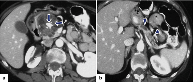
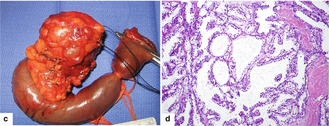
Fig. 6.26
Serous cystadenoma (microcystic type) with coarse calcifications and pancreatic duct dilatation on CT. A 72-year-old female with mild abdominal pain. CECT axial images (a, b) demonstrate a large, lobulated mass in the head of the pancreas composed of multiple small cysts and a fibrous scar. Note the coarse calcifications in the central scar (arrows), the atrophy of the pancreas, and the dilatation of the pancreatic duct distal to this cystic mass (arrowheads). The patient underwent a Whipple procedure. A photograph of the gross specimen (c) shows a lobulated mass arising from the head of the pancreas that is attached to a segment of duodenum. Histological section (d) (H&E, 20×) shows cysts of different sizes, lined by clear epithelial cells. Final pathology: serous cystadenoma microcystic type, associated with chronic pancreatitis of the distal pancreas
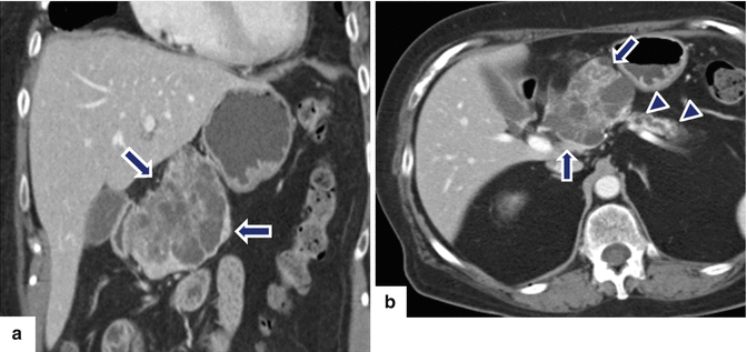
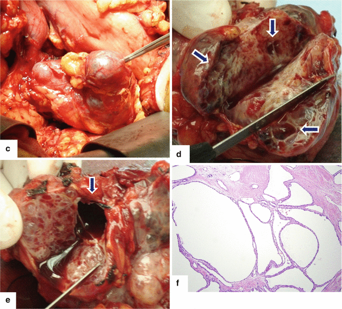
Fig. 6.27
Serous cystadenoma (microcystic type) associated with pancreatic duct dilatation on CT. A 65-year-old female with epigastric pain. CECT coronal (a) and axial (b) images show a lobulated multicystic mass (arrows) with an enhancing central scar. Note the dilatation of the pancreatic duct (arrow) and atrophy of the pancreas distal to this mass (arrowheads).Intraoperative photograph (c) shows a large, lobulated, cystic mass arising from the pancreas. Photographs of the sectioned gross specimen show a (d) cystic tumor with multiple small cysts separated by fibrous septae (arrows) and (e) a dilated pancreatic duct (arrow) distal to the cystic mass. Histological section (f) (H&E, 10×) shows a serous cystadenoma with intervening fibrotic septa. Final pathology: serous cystadenoma, microcystic type, associated with chronic pancreatitis of the distal pancreas
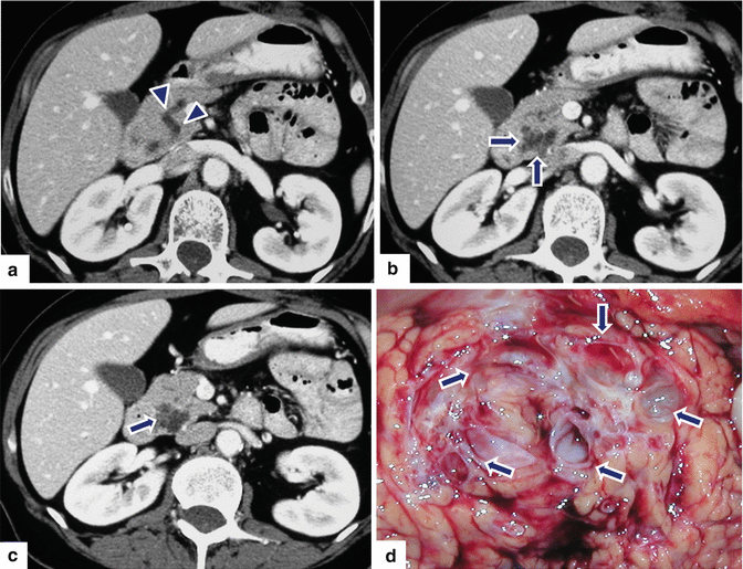
Fig. 6.28


Serous cystadenoma (microcystic type) associated with pancreatic duct dilatation on CT. A 73-year-old male with history of hepatitis B underwent a screening CT of the abdomen in which a cystic tumor was discovered. CECT axial images (a–c) demonstrate a small lobulated mass with multiple cysts and central scar involving the head of the pancreas (arrows). Note the mild dilatation of the pancreatic duct associated with this mass (a) (arrowheads). The patient underwent an endoscopic ultrasound with FNA that was non-diagnostic. A Whipple procedure was performed. A photograph of the sectioned specimen (d) shows a multicystic mass (arrows)
Related posts:
Stay updated, free articles. Join our Telegram channel
Full access? Get Clinical Tree
