KEY FACTS
Terminology
- •
Group of ovarian tumors arising from either embryonic sex cords or mesenchyme
- ○
Fibroma, thecoma, fibrothecoma
- ○
Granulosa cell tumor
- ○
Sertoli-Leydig tumor (androblastoma)
- ○
Sclerosing stromal tumor, steroid cell tumors, gynandroblastoma, and sex cord tumor with annular tubules
- ○
Imaging
- •
Ultrasound findings of sex cord-stromal tumors are diverse and nonspecific
- •
Range from small, solid tumors to large, multicystic masses
- •
Sex cord-stromal tumors are generally solid or have significant solid components
- •
Hormonally active tumors may be small and difficult to find
- •
Granulosa cell tumors
- ○
More often contain cysts with sponge-like appearance
- ○
Cysts may be complex and contain hemorrhagic fluid
- ○
- •
Fibrothecomas
- ○
Hypoechoic with posterior acoustic attenuation
- ○
May have appearance similar to uterine leiomyoma
- ○
Top Differential Diagnoses
- •
Ovarian carcinoma
- ○
Sex cord-stromal tumors less likely to have papillary projections
- ○
- •
Germ cell tumors
- ○
Much more heterogeneous with calcifications, fluid-fluid levels, etc.
- ○
Clinical Issues
- •
Symptoms related to hormone production
- •
Some are estrogen-producing tumors: Bleeding in postmenopausal patient
- •
May be associated with Meigs syndrome (triad of ovarian fibroma, ascites, pleural effusion)
Scanning Tips
- •
May mimic pedunculated fibroid; use color Doppler to look for “bridging vessels” between uterus and mass, which would suggest pedunculated fibroid over ovarian tumor
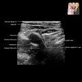
 , > expected given the hypoechoic appearance of the mass. The ovary is not identified separately, and the imaging appearance is most consistent with an ovarian fibroma.
 is homogeneously T2 dark and associated with a small claw of normal ovarian tissue
  , consistent with an ovarian fibroma.
 , which is predominately solid and vascular but also contains small cystic foci
  . This was confirmed to be a granulosa cell tumor at pathology.