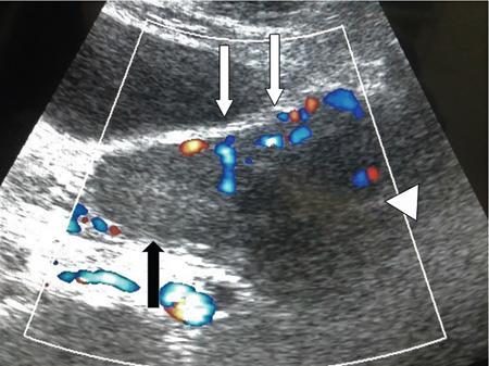
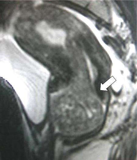
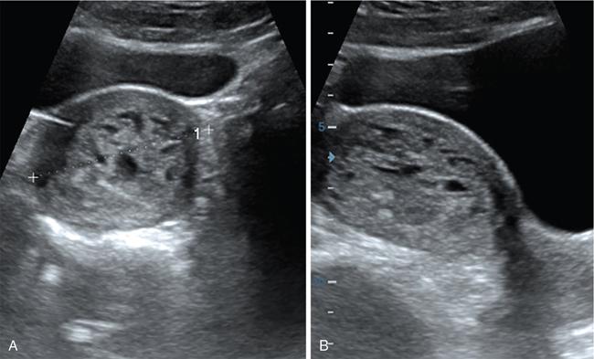
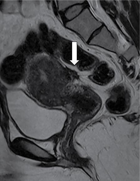
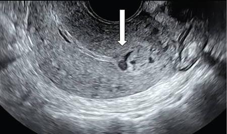
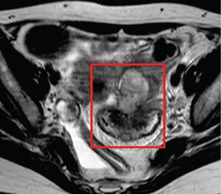
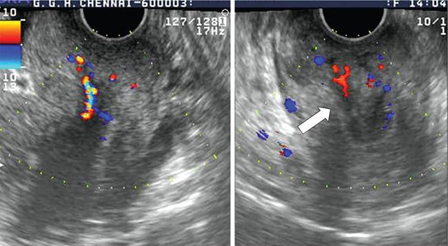
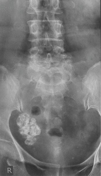
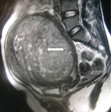
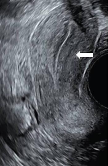
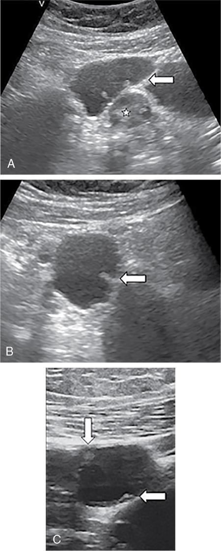
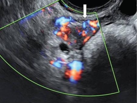
Venkatraman Indiran Bridging vessel sign Multiple vascular channels (white arrows) connecting uterus (black arrow) to an adjacent pelvic mass on imaging studies, is known as the ‘bridging vessel’ or ‘bridging vascular’ sign. Originally described on colour Doppler imaging, but also seen on computed tomography (CT) or magnetic resonance imaging (MRI). Helps in differentiating a mass of uterine origin, typically a subserosal fibroid, from other pelvic masses. Sensitivity and specificity of ‘bridging vessel sign’ in identifying subserosal fibroids range from 90% to 100%. (Best seen when subserosal fibroid is larger than 3 cm in diameter.) Ovarian masses invading the uterus may rarely produce a falsely positive bridging vessel sign. Broccoli sign Submucosal uterine leiomyoma prolapsing into the endocervical canal, along with its stalk in the uterine cavity on sagittal T2-weighted images together is called as ‘broccoli sign’ (white arrow). Submucosal uterine leiomyoma Bunch of grapes Intrauterine mass with cystic spaces without any associated foetal parts on ultrasound (white arrows) classically gives a ‘snow storm’ or ‘bunch of grapes’ appearance. Complete hydatidiform mole Hourglass sign Deep infiltrating endometriosis (DIE) affecting the posterior urinary bladder wall, usually in the midline, causes loss of its distensibility, resulting an hourglass configuration of urinary bladder. Deep infiltrating endometriosis (DIE) Hyperechoic line sign Normal endometrium surrounding the endometrial polyp on ultrasound appear as a hyperechoic rim on transvaginal ultrasound is called as ‘hyperechoic line sign’. Hyperechoic line sign represents a focal intracavitary lesion. Endometrial polyp Indian head dress sign/moose antler sign In deep infiltrating endometriosis (DIE), adhesions between the anterior wall of rectosigmoid and posterior wall of uterus, stretches the muscular layer of the bowel and creates an infiltrative nodule with parallel lines that extend from the adhesion to the rectosigmoid. This is called as ‘Indian head dress sign/moose antler sign’ (white arrow). This is seen on sagittal T2-weighted images as well as transvaginal ultrasound. Deep infiltrating endometriosis (DIE) Interstitial line sign An echogenic line that extends into the upper regions of the uterine horn and borders the margin of the intramural gestational sac is called as the ‘interstitial line sign’ (white arrow). Seen best on transabdominal/transvaginal ultrasound. Specific finding of interstitial pregnancy Mushroom cap sign Mushroom-shaped lesion which is seen displaced into the bowel lumen in patients with solid invasive endometriosis of the rectosigmoid colon on axial or sagittal T2-weighted MRI is called as ‘mushroom cap sign’ (red box and white arrow). Low signal intensity base of the mushroom corresponds to hypertrophy and fibrosis of the muscularis propria, whereas the high intensity cap represents oedematous mucosa and submucosa. Deep infiltrating endometriosis (DIE) Pedicle artery sign Feeding vessel sign Central vessel entering the endometrium from the surrounding myometrium colour Doppler imaging of the endometrium in endometrial polyps is called as pedicle artery sign (white arrow). Endometrial polyps in women with postmenopausal bleeding. (Sensitivity of 76.5%; specificity of 95.3%.) Popcorn ball calcification Dense, amorphous calcifications of uterine fibroids seen on the plain radiographs is called as ‘popcorn calcification’ (white arrow). Confluent, coarse calcification constitutes the most specific radiographic sign of a benign uterine fibroid; but only 10% or fewer of fibroids show calcification. Question mark sign of uterus The uterine corpus that is flexed backward with the fundus of the uterus facing the posterior pelvic compartment and the cervix directed anteriorly toward the urinary bladder, is called as question mark sign of uterus (white arrow). Also called as comma-shaped uterus. 93% specificity and 75% sensitivity in diagnosing adenomyosis uterus. Sliding organs’ sign When the uterus and ovaries glide freely over the posterior and anterior organs (such as rectum and urinary bladder, respectively) during real-time dynamic transvaginal sonography, it is called as ‘positive sliding organs’ sign’. In case of adhesions due to endometriosis, free gliding of organs is absent and is called as negative sliding sign. Snowstorm sign Grainy appearance with low-level homogeneous internal echoes which move slowly downward due to their viscosity seen in endometrioma on transvaginal ultrasound is called as snowstorm sign. Endometriomas Split fibre sign Presence of curvilinear T2 hypointense strands within the degenerated leiomyoma separated by the fluid accumulation and oedema is called as the ‘split fibre’ sign. Split fibre sign on T2-weighted MRI can be used to differentiate hydropic degeneration from other conditions like malignant change. Three-line sign or triple line sign or trilaminar appearance Shortly before ovulation, two additional bright linear echoes outline two hypoechoic layers of endometrium with a central hyperechoic line in the endometrium (white arrow). It is called as the ‘three-line sign’ or ‘triple line sign’ or trilaminar appearance. A triple-line pattern with a moderate endometrial thickness is associated with a good clinical outcome on in-vitro fertilization. Venetian blind shadowing Also known as ‘rain shower’ appearance Heterogeneous myometrial appearance due to hyperechoic heterotopic endometrial tissue and hypoechoic hyperplastic smooth muscle appearing as vertical linear bands of shadows on ultrasound is called as ‘Venetian blind shadowing’. Classically described in uterine adenomyosis; sometimes in uterine fibroids also. Bead on strings sign Thickened endosalpingeal longitudinal folds within a dilated fallopian tube without wall thickening appears as small mural nodules on cross-sectional images on ultrasound is called as ‘bead on strings’ appearance. Seen in chronic salpingitis Beaded tube Multiple constrictions along the course of fallopian tube may form due to scarring is seen in genitourinary tuberculosis and shows a ‘beaded’ appearance. Female genital tuberculosis Cogwheel sign Thickened endosalpingeal longitudinal folds within a dilated fallopian tube with thickened wall appears as small mural nodules on cross-sectional images across the long axis of the fallopian tube on ultrasound (white arrows) produce a characteristic ‘cogwheel’ appearance. Seen in acute salpingitis. The presence of thickened longitudinal folds is pathognomonic for hydrosalpinx. Cobblestone tube Rounded filling defects in cases of fallopian tube tuberculosis are called as the cobblestone pattern. Is an effective radiographic sign of intraluminal scarring and adhesions. Female genital tuberculosis Golf club tube Occlusion of the isthmus or fimbrial end of the fallopian tube and filling up of the tube with serous or clear fluid produces a moderate dilatation at the fimbrial end, produces a ‘golf club–like appearance’. Female genital tuberculosis Halo sign Thin radiolucency seen separating the loculated peritubal collection from the dilated tube, is known as the ‘halo sign’. This radiolucent halo represents the thickened wall of the tube. Female genital tuberculosis Incomplete septation sign Thin linear structures appearing to protrude into the tubular cystic adnexal structures, but not touching the opposite walls is called ‘incomplete septation sign’ of hydrosalpinx. Apparent septae are due to apposition of the inner walls of the dilated and folded fallopian tube. Hydrosalpinx Pipe stem tube Rigid contour of the fallopian tube seen in cases of fallopian tube tuberculosis is called as ‘pipe stem appearance’. Female genital tuberculosis Leash sign Typical eccentric leash of vessels on colour Doppler (white arrow) showing a low resistance placental type of flow on spectral Doppler in fallopian tube ectopic pregnancies is called as leash sign.
11.5: Signs in reproductive imaging
Name of the Sign
Description/Reason for the Appearance
Image With or Without Illustration
Causes and Significance

Sign
Explanation
Image
Cause
Related posts:
![]()
Stay updated, free articles. Join our Telegram channel

Full access? Get Clinical Tree

 Get Clinical Tree app for offline access


