Sinonasal Melanoma
Michelle A. Michel, MD
Key Facts
Terminology
Neural crest cell malignancy arising from melanocytes in sinonasal mucosa
Imaging
Soft tissue mass in nasal cavity > paranasal sinuses with bone destruction ± remodeling
Predilection for nasal septum, lateral nasal wall, & inferior turbinate
MR (melanotic melanoma)
↑ T1 & ↓ T2 signal results from melanin, free radicals, metal ions, and hemorrhage
T2* GRE may show “blooming” when hemorrhage present in SNM
Avidly enhances due to vascularity in SNM; enhancement may be difficult to appreciate if high pre-contrast T1 signal present
Top Differential Diagnoses
Squamous cell carcinoma
Non-Hodgkin lymphoma
Esthesioneuroblastoma
Clinical Issues
Adult with nasal stuffiness, epistaxis, & pigmented mass identified at nasal endoscopy
5th-8th decades most common
M > F
> 90% occurs in whites
Poor prognosis with 6-17% chance of 5-year survival
Mean survival 24 months
Systemic metastatic disease typically precedes death
Diagnostic Checklist
Look for mass arising in lower nasal cavity with ↑ T1 and ↓ T2 signal
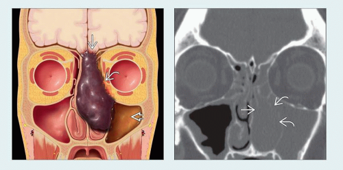 (Left) Coronal graphic shows a darkly pigmented (highly melanotic) mass centered in the nasal cavity. Invasion of the skull base
 , orbit , orbit  , and lateral nasal wall is seen, but the septum is deviated rather than invaded. Trapped secretions , and lateral nasal wall is seen, but the septum is deviated rather than invaded. Trapped secretions  are noted in the left maxillary sinus. (Right) Coronal bone CT in a patient with left nasal obstruction and epistaxis shows a mass in the left nasal cavity with erosion of the left middle turbinate are noted in the left maxillary sinus. (Right) Coronal bone CT in a patient with left nasal obstruction and epistaxis shows a mass in the left nasal cavity with erosion of the left middle turbinate  and portions of the lateral nasal wall and portions of the lateral nasal wall  . .Related posts:Stay updated, free articles. Join our Telegram channel
Full access? Get Clinical Tree
 Get Clinical Tree app for offline access

|