KEY FACTS
Terminology
- •
Microbial infection of subcutaneous tissue (cellulitis), muscle (myositis), tendon (tenosynovitis), or fascia (fasciitis)
- •
Abscess is focal fluid collection containing suppurative inflammatory material
Imaging
- •
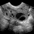
Cellulitis
- ○
Edematous, hyperechoic, hyperemic subcutaneous fat with thickened interlobular septa
- ○
- •
Infectious tenosynovitis
- ○
Synovial tendon sheath is thickened, hyperemic, and contains fluid surrounding tendon
- ○
- •
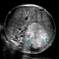
Infectious myositis
- ○
Hyperechoic, hyperemic, and indistinct, ± abscess
- ○
- •
Necrotizing fasciitis
- ○
Spreads rapidly between tissue planes
- ○
Thickened, irregular fascia with perifascial fluid
- ○
Gas is highly specific but not always present
- ○
- •
Abscess
- ○
Variable echogenicity, peripheral hyperemia, ± septation/gas
- ○
Clinical Issues
- •
Route of spread can be direct (skin), postsurgical, or hematogenous
- •
Deep soft tissue infection may require hospitalization, IV antibiotics, &/or surgery
- •
Necrotizing fascitis is life threatening and early diagnosis decreases morbidity
Scanning Tips
- •
Use wide field of view and increase depth to ensure entire area of abnormal tissue is imaged
- •
Diagnose abscess by demonstrating fluctuance and lack of color Doppler flow
- •
Note and report visible skin changes, breeches, or crepitus
- •
Assess nearby joints for effusion (septic arthritis) and adjacent bone for cortical irregularity (osteomyelitis)
 with intervening hyperechoic fat lobules
  .
.
 shows marked expansion of the tendon sheath
  with complex fluid
  as well as marked subcutaneous edema and hyperemia.
 with an intramuscular abscess
  containing layering debris.
Related posts:


Stay updated, free articles. Join our Telegram channel

Full access? Get Clinical Tree


