Disease site
Presentation
Recommended treatment
Extremity
Early stage (I)
Surgery->EBRT for +/close margin
Intermediate-advanced stage (II–III)
Surgery->post-op EBRT or pre-op EBRT->surgery, ±chemo for deep/high grade tumors
Retroperi-toneal
Resectable
Surgery + IORT- >post-op EBRT or pre-op EBRT->Surgery +IORT
GIST
Resectable
Surgery ± imatinib
Unresectable
Imatinib- >re-eval±surgery
Desmoid
Resectable
Surgery±EBRT for +margin
Unresectable
EBRT, chemo
Metastatic (stage IV)
Chest, abdominal, or pelvic oligometastases
Surgical metastatectomy, SBRT, systemic therapy
Spinal metastases
Surgical resection/stabilization, EBRT/ SBRT
Diffuse metastases
Systemic therapy, EBRT/ SBRT for palliation of selected involved sites
Radiosurgical Technique
Dose and fractionation directed by adjacent normal tissue RT toxicity constraints.
Simulation and Treatment Planning
If biopsy or resection performed, request fiducial marker placement.
Prefer fine-cut (<5 mm) treatment planning CT ± contrast with 4DCT to define ITV for thoracic or upper abdominal metastases.
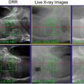
Immobilization with body frame and/or fiducial , lesion, or vertebral element tracking.
Abdominal compression and/or respiratory gating may be employed to reduce lesion motion associated with diaphragmatic excursion during breathing.
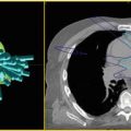
Image fusion with diagnostic CT, MRI , myelogram , and/or PET for target delineation as appropriate.
GTV /iGTV = lesion as defined by CT- or MRI -based imaging , with contrast as available. Lung windowing should be used for pulmonary oligometastases.
CTV /ITV = GTV /iGTV + 0–10 mm (CTV/ITV=GTV/iGTV for pulmonary lesions).
PTV = CTV /ITV + 3–5 mm (smaller margins with intrafraction image guidance and/or motion management).
Image guidance with orthogonal kV and/or cone beam CT for daily treatment delivery.
Dose Prescription
Central lung oligometastases.
10 Gy × 5 fx.
Peripheral lung oligometastases.
25–34 Gy × 1 fx, 18 Gy × 3 fx, 12 Gy × 4 fx, 10 Gy × 5 fx.
Abdominal and pelvic oligometastases.
6–8 Gy × 5 fx.
Vertebral spine metastases.
18–24 Gy × 1 fx, 8 Gy × 3 fx, 6 Gy × 5 fx.
Toxicities and Management
EBRT late radiation morbidities include decreased range of motion secondary to fibrosis at primary site, lymphedema with circumferential treatment of extremities, and low risk of secondary malignancy .
SBRT toxicity related to dose and volume of treated adjacent normal tissues.
Risk of lung injury for pulmonary metastases (see Chap. 7).
Risk of liver , adrenal, renal, and bowel toxicity for abdominal metastases.
Nausea most common acute toxicity for abdominal SBRT , often responsive to short-term antiemetic pharmacotherapy .
Acute pain flare and risk of late myelopathy for spinal metastases.
Recommended Follow-Up
Exam with functional status , MRI of primary, CT chest every 3 months × 2 years, then every 4 months × 1 year, then every 6 months × 2 years.
CT imaging of treated oligometastatic site every 3 months × 1 year.
Consider bone scan, MRI , or PET , if clinically indicated.
Evidence
There is lack of randomized prospective data on use of SBRT approaches in primary, recurrent, and metastatic disease.
Primary STS
While there is limited data regarding SBRT in management of primary STS , there is evidence of efficacy of short-course adjuvant hypofractionated RT delivered via brachytherapy , as well as improved outcomes with reduced treatment-volume IGRT and IMRT techniques.
Itami et al. (2010): Retrospective series of 25 primary STS patients treated postoperatively with HDR monotherapy, 36 Gy in 6 fx in 3 days b.i.d. LC 78.2 % at 5 years, but up to 93.3 % for patients with negative margins and no prior surgical resections. Complication rate of 11.5 % >grade 2.
Petera et al. (2010): Retrospective series of 45 primary or recurrent STS patients treated post-operatively with HDR monotherapy (30–54 Gy, 3 Gy b.i.d fx) vs. HDR (15–30 Gy, 3 Gy b.i.d. fx)+ EBRT (40–50 Gy at 1.8–2 Gy fx). LC 74 % and OS 70 % at 5 years. LC better for primary tumors (100 %) and for patients treated with combination HDR + EBRT vs. HDR monotherapy (OR 0.2, p = 0.04).
Dickie et al. (2010) and Wang et al. (2011): Two phase II studies of increased conformity of treatment volumes employing image-guided preoperative IMRT suggest improved rates of wound complications and late morbidities including fibrosis , joint stiffness , and edema .
Alektiar et al. (2008). Retrospective series of 41 STS patients treated with IMRT in pre- and postoperative setting with increased bone sparing as compared with prior 3DCRT techniques. Favorable 5-year LC of 94 %.
Soyfer et al. (2013): Series of 21 elderly patients with median age 80 underwent post-operative hypofractionated EBRT 39–48 Gy in 13–16 fx. LC 86 % at median follow-up of 26 months. Three patients had LR, all with <3 mm surgical margins. Three patients noted with late grade 2–3 toxicity.
Levine et al. (2009): Retrospective series of primary and metastatic spinal sarcoma s treated with SBRT , including 14 primary, largely STS , spinal sarcomas. Seven patients were treated definitively with SBRT (24–35 Gy in 3–5 fx) with OS 100 % and LR 29 % at mean follow up of 33 months. Seven patients received adjuvant SBRT (3 preoperatively, 4 postoperatively for +margins), treated with 25–30 Gy in 2–5 fx. Two of three preoperatively treated patients died of recurrent disease. OS 100 % for postoperatively treated patients with median follow-up of 43.5 months. There was one instance of severe late toxicity involving rectal tumor cavity fistula in a definitively SBRT-treated patient.
Metastatic STS
Surgical Ablation
Potential survival benefit for ablative treatment of oligometastatic disease suggested by multiple surgical series.
Billingsley et al. (1999): MSKCC series of 719 patients with STS pulmonary metastases. MS 33 months for patients receiving complete metastatectomy vs. 11 months for those receiving non-operative therapy.
van Geel et al. (1996): Retrospective multi-institutional series of 255 patients with pulmonary STS metastases. OS 42 and 35 % at 3 and 5 years, respectively. Young age (<40), R0 resection, and low/int grade tumors associated with better OS.
Porter et al. (2004): Comparative effectiveness study of surgical metastatectomy vs. systemic chemotherapy for treatment of pulmonary STS metastases. Despite favorable assumptions of benefit of chemotherapy, surgical ablative therapy was deemed a significantly more cost-effective management approach.
DeMatteo et al. (2001): 331 patients treated at MSKCC for STS liver metastases . 56 patients underwent R0 or R1 gross resection of hepatic metastases, with MS 39 months vs. 12 months for those who did not undergo complete or any resection independent of adjuvant systemic therapy.
Marudanayagam et al. (2011): Retrospective series of 36 patients who underwent hepatic resection for oligometastatic STS . OS from metastatectomy was 90.3 % (1 year), 48.0 % (3 years), 31.8 % (5 years). Poor survival associated with high-grade tumors, primary leiomyosarcoma , and positive resection margin of liver metastasis .
Radiotherapy
Merrell et al. (2014): Retrospective series of 21 patients with metastatic STS receiving SBRT . Median dose 50 Gy in 5 fx (lung), 24 Gy in 1 fx (bone ), 42.5 Gy in 5 fx (liver ), and 40 Gy in 4 fx (soft tissue). LC was 94 %(12 months), 83 % (24 months), and 83 % (48 months). Most frequent toxicities were of low grade, including acute pain flare and nausea , and late cough .Related posts:

Stay updated, free articles. Join our Telegram channel

Full access? Get Clinical Tree