Fig. 9.1
Solid pseudopapillary tumor of the pancreas gross appearance in situ. Intraoperative photographs of two patients show (a) a large, ovoid, encapsulated mass with smooth surface in the head of the pancreas and (b) a yellowish slightly lobulated mass in the body and tail of the pancreas (arrows)
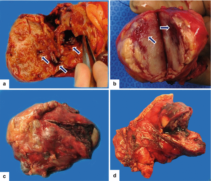
Fig. 9.2
Solid pseudopapillary tumor of the pancreas macroscopic appearance. Macroscopic images of four different specimens that have been bisected demonstrate (a) a partially cystic mass with areas of hemorrhage (arrows), (b) a round, solid, white-tan, soft mass with focal areas of hemorrhage (arrows), and (c, d) round and soft, encapsulated hemorrhagic masses
Soft, round or ovoid in shape
Tan-brown, well-circumscribed mass
Regions of necrosis, hemorrhage, and cystic degeneration
Thick fibrous capsule
Calcifications may be present
Average size: 7–10 cm
9.3.2 Microscopic Appearance
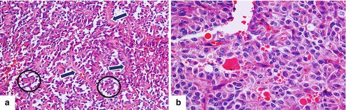
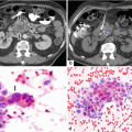
Fig. 9.3
Solid pseudopapillary tumor of the pancreas microscopic appearance. Histology slides (a) show thin fibrovascular cores (arrows) lined by epithelial cells, forming pseudopapillae. The circles highlight the detached degenerating epithelial cells (H&E, 10×). In more solid areas of the tumor, (b) the cells remain cohesive and can produce hyaline eosinophilic globules, which are usually positive for PAS stain (H&E, 20×)
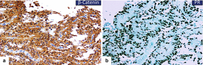
Fig. 9.4
Solid pseudopapillary tumor of the pancreas. Immunohistochemistry shows nuclear positivity for (a) β-catenin (10×) and (b) progesterone receptor (PR) (20×) in the tumor cells
Thin fibrovascular core lined by bland epithelial cells with eosinophilic cytoplasm
Intracytoplasmic PAS + hyaline globules are present
These tumors are positive for:
β-catenin, neuron-specific enolase (NSE), alpha-1-antytrypsin, CD10, CD56, synaptophysin, and progesterone receptor (PR)
These tumors are negative for:
Chromogranin, epithelial membrane antigen (EMA), carcinoembryonic antigen (CEA), trypsin, chymotrypsin, and lipase
9.4 Clinical Presentation
Abdominal pain (most common)
Asymptomatic, incidentally discovered (physical examination or imaging)
Abdominal fullness
Palpable mass
Early satiety
Nausea and vomiting
Pancreatitis
Hematemesis
Jaundice
9.5 Laboratory Evaluation
Nonspecific
Rare: hyperbilirubinemia or mild elevation of amylase or lipase levels
CEA and CA19-9 markers are negative
9.6 Imaging
Preferred imaging modalities: computed tomography and magnetic resonance
In equivocal cases, endoscopic ultrasound is preferred
9.6.1 Ultrasound (US)
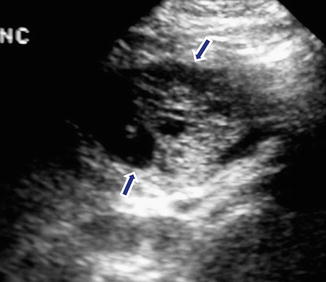
Fig. 9.5
Solid pseudopapillary tumor on ultrasound. An 18-year-old female with left upper quadrant pain. Sagittal view shows an ovoid complex mass with solid and cystic components in the tail of the pancreas (arrows)
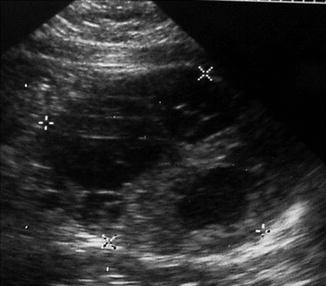
Fig. 9.6
Complex appearance of a solid pseudopapillary tumor on ultrasound. A 23-year-old female patient with epigastric fullness. Transverse view reveals a complex mass with solid and cystic components in the head of the pancreas (crosses)
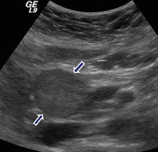
Fig. 9.7
Solid pseudopapillary tumor of the pancreas on ultrasound. A 16-year-old female with epigastric pain. Transverse view demonstrates a large, solid, round mass in the head of the pancreas (arrows)
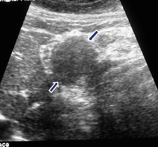
Fig. 9.8
Solid pseudopapillary tumor of the pancreas. An 18-year-old patient with epigastric pain. Sagittal view demonstrates a well-circumscribed, solid, ovoid mass in the body of the pancreas (arrows)
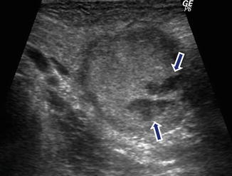
Fig. 9.9
Partially necrotic solid pseudopapillary tumor of the pancreas. A 28-year-old female with left upper quadrant pain. Intraoperative ultrasound transverse view reveals a large mass mostly solid with cystic components in the tail of the pancreas (arrows)
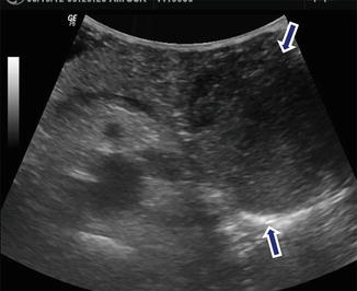
Fig. 9.10
Heterogeneous solid pseudopapillary tumor of the pancreas on ultrasound. A 38-year-old female with left upper quadrant discomfort. Transverse view shows a mass with heterogeneous echogenicity involving the tail of the pancreas (arrows)
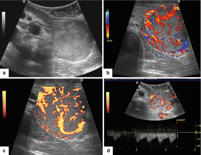
Fig. 9.11
Hypervascular solid pseudopapillary tumor on intraoperative ultrasound. A 29-year-old female with left upper quadrant fullness. Gray scale (a), color (b), and power Doppler (c, d) transverse views demonstrate a round, solid hypervascular mass in the tail of the pancreas
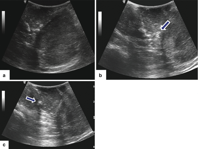
Fig. 9.12
Solid pseudopapillary tumor on intraoperative ultrasound (IOUS). A 60-year-old female with a pancreatic mass found incidentally on a screening ultrasound to evaluate the liver. Transverse views (a–c) demonstrate a large bilobulated solid mass with coarse calcifications (arrows) on the tail of the pancreas
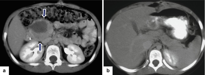
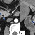
Fig. 9.13
Necrotic solid pseudopapillary tumor of the pancreas on CT. A 19-year-old female with mild epigastric pain for 1 month. CECT axial images demonstrate a round, pancreatic necrotic mass in the head of the pancreas (a) (arrows). Note the absence of dilatation of the pancreatic and common bile ducts (b)
Encapsulated masses
Hyperechoic
Hypoechoic
Complex mass (solid and cystic components, internal septations)
Central or peripheral echogenic foci (calcifications)
Color Doppler: avascular or hypervascular masses
9.6.2 Contrast-Enhanced Computed Tomography (CECT) (Figs. 9.13–9.35)
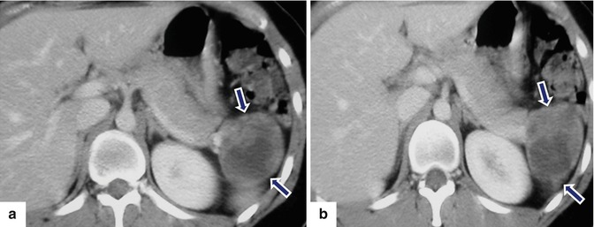
Fig. 9.14
Necrotic solid pseudopapillary tumor of the pancreas on CT. A 24-year-old female with left upper quadrant pain. CECT axial images (a, b) show a complex mass with solid and cystic components involving the tail of the pancreas (arrows)
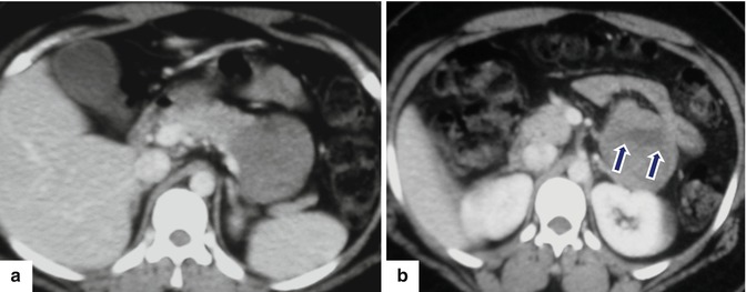
Fig. 9.15
Partially necrotic solid pseudopapillary tumor of the pancreas on CECT. A 33-year-old female with abdominal discomfort. CECT (a, b) axial images show a mostly solid mass with central necrosis (arrows)
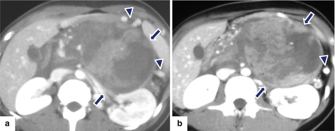
Fig. 9.16
Complex large solid pseudopapillary tumor of the pancreas on CT. An 11-year-old female with upper abdomen fullness. Contrast-enhanced CT axial (a, b) images demonstrates a large, complex mass, mostly solid with cystic components in the body and tail of the pancreas displacing the stomach anteriorly (arrows) and associated with occlusion of the splenic vein. Note the prominent short gastric veins (arrowheads)
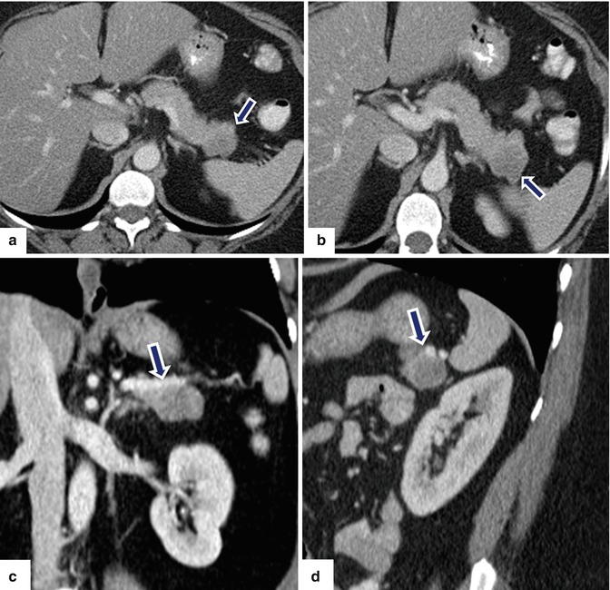
Fig. 9.17
Solid pseudopapillary tumor of the pancreas on CT. A 56-year-old female with a pancreatic tumor found incidentally on a CT performed for evaluation of nephrolithiasis. The patient underwent an endoscopic ultrasound with fine-needle aspiration of the mass. Cytologic examination showed a neoplastic process with neuroendocrine features. CECT axial (a, b), coronal (c), and sagittal (d) images show a lobulated homogeneous hypodense solid mass involving the tail of the pancreas (arrows). The patient underwent a distal pancreatectomy and splenectomy. Final pathology: solid pseudopapillary tumor of the pancreas
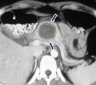
Fig. 9.18
Homogeneous pseudopapillary tumor of the pancreas on CT. A 29-year-old female with mild epigastric pain. CECT axial image shows a round hypodense mass in the body of the pancreas (arrows)
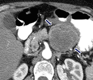
Fig. 9.19


Heterogeneous pseudopapillary tumor of the pancreas on CT. A 38-year-old female with a pancreatic mass found incidentally on a workup for a renal mass. CECT axial image reveals a heterogeneous low-density mass in the body of the pancreas (arrows)
Related posts:
Stay updated, free articles. Join our Telegram channel
Full access? Get Clinical Tree
