Spectroscopy Of The Breast
Peter Stanwell, PhD
Carolyn Mountford, D.Phil
Terminology
Abbreviations
Magnetic resonance spectroscopy (MRS)
Definitions
Detectable biochemicals are distinguished by their resonance frequency, line shape, line width, phase, and peak area
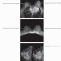
Anatomy-Based Imaging Issues
Overview
MRS success depends on
Appropriate pre-acquisition set up
Acquisition protocol
Post-processing
Shimming for MRS acquisition is essential to overcome magnetic field inhomogeneities
MRS pitfalls
B0 inhomogeneities are often dominant factor limiting successful MRS
Some breast coils are not optimized for spectroscopy
Presence of biopsy clips may affect shimming
Some gadolinium-based MR agents have been shown to affect breast MRS
Lipids
Localized shimming more difficult due to abundance of adipose tissue with different susceptibility from surrounding glandular (and malignant) tissue
Impact of unwanted lipid signals can be reduced by decreasing amplitude of lipid signal
Techniques for lipid suppression exist for use in MRS
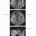
Spectroscopy Acquisition Protocols
Data Acquisition
Parameters set during pre-scan include power calibrations, frequency adjustment, shimming, and water suppression adjustment
Shimming = process by which B0 field is made as homogeneous as possible, typically using linear x-, y-, and z-gradient coils
Water suppression = process by which water signal is suppressed to allow detection of important metabolites
Spectroscopy Processing
Processing Steps
Water referencing
Aligns spectral resonances against water set at 4.74 ppm
Allows for correction of resonance distortions using water signal as reference
Apodization
In clinical application, this mathematical function is used to improve SNR
Zero filling
Method of improving spectral resolution
Fourier transformation
Converts time domain signal into frequency domain
Phase correction
Allows for correction of distortions in final spectrum
Baseline correction
Allows for correction of baseline errors in final spectrum
Vertical display may need to be increased to see resonances between 3.00 and 3.40 ppm
Spectra Interpretation
Malignant vs. Benign
Key metabolite in breast spectroscopy is choline resonance at 3.23 ppm
Malignant breast cancers show resonance at 3.23 ppm
Benign breast lesions show resonance at 3.28 ppm
This resonance is reported in breast tissue in lactating and other healthy women
Differential Diagnosis
Invasive Tumor
Early single series showed sensitivity of 100% and specificity of 88%
Selected References
Related posts:


Stay updated, free articles. Join our Telegram channel

Full access? Get Clinical Tree


