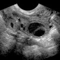
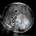
KEY FACTS
Terminology
- •
Open spina bifida (bone and skin defect ± sac)
- ○
Meningomyelocele: Sac + neural elements
- ○
Meningocele: Sac with fluid only
- ○
Myeloschisis: No sac (cord directly exposed)
- ○
- •
Closed spina bifida: Skin-covered defect
Imaging
- •
Level: Lumbosacral > thoracic > cervical
- •
Transverse view best for seeing dorsal element divergence
- •
Longitudinal views and 3D best for evaluating level
- •
Myelomeningocele: Complex cystic mass (most common)
- •
Meningocele: Anechoic cystic mass
- •
20% are myeloschisis (subtle): No sac, cord is part of defect
- •
Chiari 2 brain findings with open spina bifida
- ○
Cerebellar compression, loss of cisterna magna
- ○
- •
Tethered cord (cord seen at level of defect)
- •
Associated anomalies: Club feet, scoliosis
- •
Closed spina bifida: Chiari 2 absent or minimal
Clinical Issues
- •
↑ maternal serum α-fetoprotein (msAFP)
- •
Associated maternal folate deficiency
- ○
From maternal metabolic gene defect
- ○
Preconception high-dose folic acid for next pregnancy
- ○
- •
4% aneuploidy rate when isolated (14% if other anomalies)
- •
Prognosis depends on level and presence of hydrocephalus
- ○
80% require ventricular shunting or bypass of hindbrain compressive obstructive hydrocephalus (from Chiari 2)
- ○
Only 17% with normal continence
- ○
Scanning Tips
- •
Look carefully at spine if cisterna magna is compressed (Chiari 2 is almost never isolated)
- •
Use last rib to determine T12 and count inferiorly to determine level of spina bifida
- •
If sac covering looks thick, follow it laterally to see if it is contiguous with skin (closed defect)
 contain only cerebrospinal fluid, while myelomeningoceles
  also contain neural elements (most common). The neural tube defect is uncovered (no sac) in myeloschisis
  and contains the spinal cord or nerve roots. Also, the cord is tethered to the defect.
 , which communicates with the spinal canal
  . The finding is classic for open spina bifida with sac.
 ) wraps around the midbrain
  , taking on the typical banana shape described with Chiari 2 malformation.
 extending through splayed dorsal spine ossification centers (arches)
  and into the myelomeningocele sac
  . Chiari 2 is highly associated with open spina bifida.
Stay updated, free articles. Join our Telegram channel

Full access? Get Clinical Tree


