Fig. 41.1
Magnetic Resonance Imaging sagittal T2 through the cervical spine demonstrating traumatic disc protrusions (solid black arrow), long segment cord edema (open arrow), focal cord hemorrhage, and extensive posterior cervical soft tissue edema (white arrow)
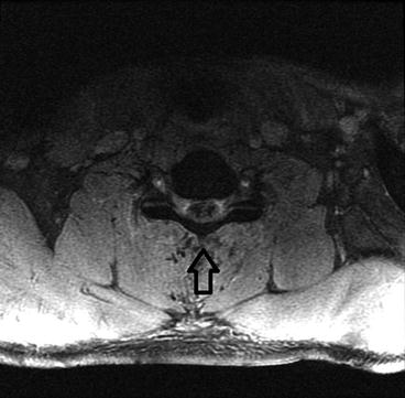
Fig. 41.2


Magnetic Resonance Imaging axial Multiple Echo Recombined Gradient Echo (MERGE) 2D through the cervical spine demonstrating gradient susceptibility in the cervical cord confirming hemorrhage (open arrow)
Related posts:


Stay updated, free articles. Join our Telegram channel
Full access? Get Clinical Tree
