2
Spine
There will be ten questions on imaging procedures for the spine in the advanced-level examination for MRI. These will relate to the following areas
- Cervical
- Thoracic
- Lumbo-sacral
Q41 Most spine imaging is performed with the use of:
| (a) | A surface/local coil |  |
| (b) | ECG gating |  |
| (c) | Respiratory compensation |  |
| (d) | Peripheral gating |  |
Q42 In patients that have undergone surgery for a herniated disk in the lumbar spine, contrast enhancement can be used to distinguish recurrent disk from post-operative scar because:
| (a) | Post-operative scar never enhances and recurrent disk does enhance |  |
| (b) | Post-operative scar enhances and recurrent disk does not |  |
| (c) | Disk enhances more slowly than post-operative scar |  |
| (d) | Neither scar nor disk enhance |  |
Q43 For optimal imaging of the cervical spine, patient positioning and local coil placement are:
| (a) | Supine/under the neck to include from C1 to C7 |  |
| (b) | Supine/on top of the neck to include from C1 to C7 |  |
| (c) | Supine/beside the neck to include from C1 to C7 |  |
| (d) | Prone/on top of the neck to include from C1 to C7 |  |
Q44 On a 24 cm FOV, sagittal T-spine image that demonstrates a cord compression, the vertebral level can be determined by:
| (a) | Using the xyphoid as a landmark and counting up from T12 |  |
| (b) | Using the sternal notch as a landmark and counting down from T1 |  |
| (c) | Using a large FOV localizer and counting down from C2 |  |
| (d) | Using lead markers to mark T12 and T1 on large FOV images |  |
Q45 In lumber spine imaging, images acquired directly through intervertebral disk spaces can be acquired in the:
| (a) | Axial plane |  |
| (b) | Sagittal plane |  |
| (c) | Coronal plane |  |
| (d) | Oblique plane |  |
Q46 On T1 weighted images of the spine, the CSF appears:
| (a) | Hyperintense to the spinal cord |  |
| (b) | Hypointense to the spinal cord |  |
| (c) | Isointense to the spinal cord |  |
| (d) | a and c |  |
Q47 The conus and the cauda equina in adult patients are best demonstrated by a:
| (a) | Sagittal image of the cervical spine |  |
| (b) | Sagittal image of the thoracic spine |  |
| (c) | Sagittal image of the lumbar spine |  |
| (d) | Coronal image of the thoracic spine |  |
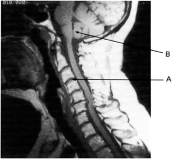
Q48 Image 4 was acquired in the:
| (a) | Axial imaging plane |  |
| (b) | Sagittal imaging plane |  |
| (c) | Coronal imaging plane |  |
| (d) | Off-axis (oblique) imaging plane |  |
Q49 Image 4 is an example of:
| (a) | A T1 weighted image |  |
| (b) | A T2 weighted image |  |
| (c) | A spin (proton) density weighted image |  |
| (d) | A T2* weighted image |  |
| (e) | All of the above |  |
Q50
Related posts:
Stay updated, free articles. Join our Telegram channel

Full access? Get Clinical Tree


