165 Spleen, kidney
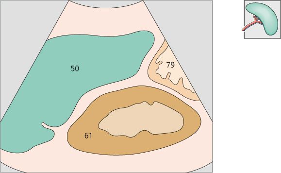
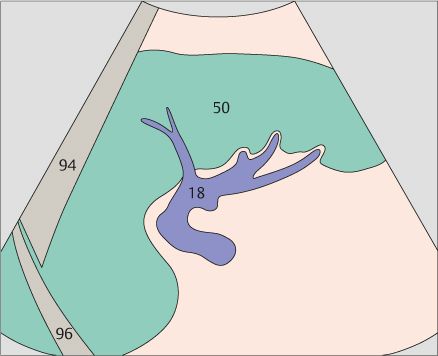
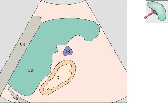
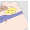
166 Splenic hilum, splenic vein
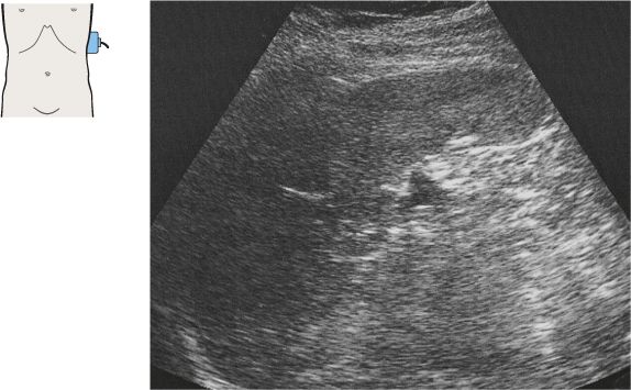
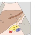
The spleen is identified in the longitudinal flank scan as a rounded triangle between the upper renal pole and the diaphragm.
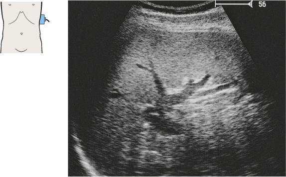
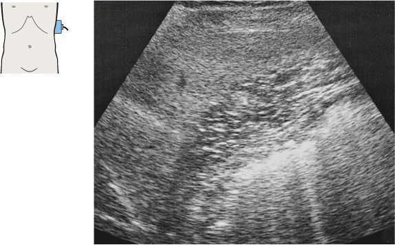
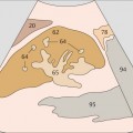
A flank scan at the level of the hilum displays the spleen in its greatest longitudinal dimension.
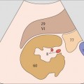
167 Spleen, stomach
168 Spleen, stomach
Stay updated, free articles. Join our Telegram channel

Full access? Get Clinical Tree


