Chapter 5. Spleen
Patient Preparation
• No preparation is required, although if it is included in a complete abdomen examination, the patient may be fasting.
Equipment and Technical Factors
• Curved linear multihertz transducer is needed; sector/vector transducer may be required for intercostal imaging.
• The spleen demonstrates a very homogenous echotexture and midlevel echogenicity that is isoechoic to hypoechoic to the liver; the blood vessels are generally only noted at the hilum.
• Color Doppler imaging may confirm the direction of flow in the splenic vessels.
Imaging Protocol
• A variety of transducer placements (scan planes) may be used to obtain diagnostic images of the spleen; therefore, each image must be labeled accurately for scan plane and anatomy demonstrated.
• Longitudinal axis images through the medial, mid (hilum), and lateral aspects of the spleen are obtained; if the coronal plane is used, then the anterior, mid, and inferior aspects are documented.
• Transverse axis images through the superior, mid (hilum), and inferior aspects of the spleen are obtained; the transverse lateral scan plane may be used to obtain these images.
Sonographic Measurements
The spleen is variable in shape.
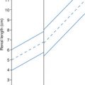
• Length: <13 cm as measured from superior to inferior pole; 7.0−9.0 cm if measured from the diaphragm to inferior tip (range 5.5−14.0 cm)
• Width (at hilum): variable (range: 6.0−12.0 cm)
• Depth (AP): <6.0 cm (range 3.0−8.0 cm)
• Volume Index (adult): 8−34 SVI = Length × Height × Thickness/27
Note: the depth measurement may be done in either the longitudinal or transverse axis images; if the coronal plane is used for the longitudinal axis of the spleen, the depth cannot be measured in the same image.
| Spleen | |||
|---|---|---|---|
| Sonographic Finding(s) | Clinical Presentation | Differential Diagnosis | Next Step |
Inferior border of spleen extends past mid left kidney or the spleen appears extremely prominent Echogenicity may be normal or decreased | Associated with one or more of the following: Heart failure Portal hypertension Leukemia
Related posts:Stay updated, free articles. Join our Telegram channel
Full access? Get Clinical Tree


| ||