Splenic Embolization in Nontraumatized Patients


Splenic Embolization in Nontraumatized Patients


Robert F. Dondelinger


Indications


Arterial embolization of the spleen may have two different objectives: interruption of arterial flow to the splenic artery or one of its branches, or ablation of splenic tissue by infarction. Depending on the aim of the procedure, splenic embolization may be considered an alternative to either splenectomy or ligation of the splenic artery. A typical indication for interruption of flow in the splenic artery or one of its branches is posttraumatic hemorrhage.


Splenic artery embolization has also been advocated to treat bleeding due to ruptured gastroesophageal varices, but portal hypertension has not been proven to be significantly influenced in all cases by arterial splenic embolization when portal pressure was recorded.


Catheter-mediated infarction of splenic tissue may be advocated as an alternative to splenectomy in various pathologic conditions. The volume of the spleen may be subnormal, but in the vast majority of cases, splenomegaly is obvious. Because intrasplenic arterial vascularization is represented by terminal vessels, efficient infarction of splenic tissue is regularly achieved after embolization of the intrasplenic branches.


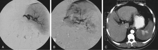
Hypersplenism due to portal hypertension is certainly the primary indication for embolization of arterial splenic tissue.1,2 Causes of hypersplenism vary. Most patients have alcoholic or postnecrotic liver cirrhosis, with a platelet count of less than 60,000/mm3 and at least one major episode of bleeding from ruptured gastroesophageal varices. Embolization of the spleen has the advantage of retaining the organ in place, and a distal splenorenal shunt is still possible after correction of the anemia and thrombocytopenia.


Children with gastroesophageal bleeding as a sign of hypersplenism secondary to Gaucher disease, atresia of the intrahepatic bile ducts, or portal vein thrombosis have also been treated by splenic embolization.


Splenic embolization is also indicated in patients being maintained on hemodialysis who have pancytopenia and splenomegaly, and in renal transplant patients in whom intolerance to immunosuppressive medications after renal transplantation had to be corrected.3


Thalassemia is also responsible for progressive hypersplenism, which requires an increase in the number of blood transfusions.4,5 Partial splenic embolization is a valid alternative to splenectomy in this group of patients, in whom the risk for infections and lethal complications is particularly high after splenectomy.


Idiopathic thrombocytopenic purpura may also be corrected by splenic embolization.6,7 The procedure is well tolerated in these patients because the spleen is of normal volume or only moderately enlarged. The physiopathologic mechanism of correction of thrombocytopenia has not been completely elucidated. Infarction of splenic tissue probably decreases sequestration of platelets in the spleen and reduces intrasplenic production of autoantibodies directed against platelets.


Splenic embolization has been used in a variety of other pathologic conditions, such as splenic lymphoma, chronic lymphatic leukemia, myeloid leukemia, myelofibrosis, hairy cell leukemia, polycythemia vera, hereditary spherocytosis, autoimmune hemolytic anemia, idiopathic hypersplenism, and Felty syndrome, as well as in patients with hypersplenism and cytopenia induced by anticancer chemotherapy.811


Preoperative partial splenic embolization has also been advocated before laparoscopic splenectomy,12,13 in a tumorous spleen when correction of hypersplenism and reduction of tumor mass are expected, and in patients with hypersplenism, those at high operative risk, and those who refuse blood transfusion (Jehovah’s Witnesses).14


Technique


Anatomy and Approach


For anatomy of the celiac trunk and splenic vessels, refer to Chapter 52.


Splenic embolization is performed after selective catheterization of the splenic artery via a femoral or (more rarely) axillary or brachial approach.


Technical Aspects


Antibiotic Prophylaxis


To avoid septic complications after splenic embolization, the patient undergoes antiseptic preparation before the procedure. A bath of povidone-iodine is given the day before embolization and the puncture site is prepared. Systemic antibiotics are administered starting on the day of embolization according to different protocols, which should be adapted to local preference:


Catheterization Technique


In the vast majority of cases, standard preshaped 5F Cobra or Sidewinder catheters allow optimal catheterization of the splenic artery, with the tip of the catheter being advanced a sufficient distance from the ostium into the splenic artery. It is recommended that the splenic artery be catheterized as distally as possible to reduce any possible reflux of embolic material into the gastric and pancreatic arteries originating from the splenic artery, into other branches of the celiac artery, or into the abdominal aorta. A coaxial technique may be used in a tortuous splenic artery to optimize distal catheterization, but antegrade flow should be maintained in the splenic artery during embolization to prevent reflux of embolization agents. The risk of inadvertent embolization of caudal pancreatic arteries by migrant embolic material is not clinically significant when large particles are used as the embolization material. A branch of the splenic artery that usually originates proximal to the hilar branches and is directed to the upper pole of the spleen may be overlooked by catheterization that is too distal.


Embolization Technique and Material


Gelatin pledgets (1 × 1 × 5 mm) prepared by the operator during the procedure are the most commonly used material for splenic embolization. Use of Gelfoam powder may be associated with potential complications, such as pancreatitis and necrosis of the gastric wall secondary to thrombosis of many small terminal arteries. Particles of different size are injected, smaller particles first to block the most distal intrasplenic arteries, and large particles at the end to block the arteries at the splenic hilum. Formation of a plug of gelatin particles early in the procedure has to be avoided because it may preclude further correct embolization of the distal branches. Between 20 and 40 gelatin pledgets are necessary, depending on the volume of the spleen. Gelfoam pledgets are soaked in an antibiotic solution before injection.


The use of large steel coils lodged in the splenic artery does not induce infarction of splenic tissue. Coils are a safe embolization material, but the best long-term positive results are obtained when the coils are lodged in branches at the splenic hilum.


The extent of infarction has to be assessed during the procedure. When a significant reduction in flow of contrast medium is observed in the splenic artery, embolization is stopped. At that moment, the extent of infarction generally corresponds to 70% to 80% of the splenic volume (Fig. 72-1). Angiography can be performed during the procedure to assess the progression and extent of embolization. Angiographic catheter exchange is avoided during the procedure to prevent septic complications.


Dec 23, 2015 | Posted by in INTERVENTIONAL RADIOLOGY | Comments Off on Splenic Embolization in Nontraumatized Patients

Full access? Get Clinical Tree

Get Clinical Tree app for offline access