, Maria Custódia Machado Ribeiro2 and Bruno Beber Machado3
(1)
Hospital da Criança de Brasília – Jose Alencar, Clínica Vila Rica, Brazília, Brazil
(2)
Hospital da Criança de Brasília – Jose Alencar, Hospital de Base do Distrito Federal, Brasília, Brazil
(3)
Clínica Radiológica Med Imagem, Unimed Sul Capixaba, Santa Casa de Misericordia de, Cachoeiro de Itapemirim, Brazil
Abstract
Children and adolescents have become more and more involved in sports practice, whether competitive or purely recreational. The association of skeletal immaturity and intense physical activity renders this group age especially prone to musculoskeletal lesions. Generally speaking, chronic lesions are more common in pediatric athletes than those related to acute trauma. The latter, caused by the sudden action of high-energy forces, usually do not pose a diagnostic dilemma, while chronic osteoarticular lesions caused by repetitive trauma of low intensity may mimic articular diseases. This chapter will discuss some of the most frequent sports-related lesions, such as the different forms of apophysitis, osteochondritis dissecans, stress-related physeal lesions, and stress fractures.
10.1 Introduction
Children and adolescents have become more and more involved in sports practice, whether competitive or purely recreational. The association of skeletal immaturity and intense physical activity renders this group age especially prone to musculoskeletal lesions. Generally speaking, chronic lesions are more common in pediatric athletes than those related to acute trauma. The latter, caused by the sudden action of high-energy forces, usually do not pose a diagnostic dilemma, while chronic osteoarticular lesions caused by repetitive trauma of low intensity may mimic articular diseases. This chapter will discuss some of the most frequent sports-related lesions, such as the different forms of apophysitis, osteochondritis dissecans, stress-related physeal lesions, and stress fractures.
10.2 Apophysitis
An apophysis is an ossification center that – unlike the epiphysis – does not contribute to longitudinal bone growth; instead, it serves as an insertion site for tendons, being especially subjected to traction forces and anomalous stress. In addition, the apophyseal physes are inherently weaker and more vulnerable to traumatic lesions than the adjacent bones and tendons. These lesions may be represented by acute apophyseal avulsions at the level of the growth plate, or by chronic apophysitis, which is an inflammatory process typical of skeletally immature athletes related to excessive traction and microavulsions at the bone/cartilage interface. The latter is commonly found during growth spurts, and it may be considered a non-displaced transphyseal stress fracture (Salter-Harris type I – see Chap. 9). Patients with chronic apophysitis present insidious local pain that worsens with physical activity, as well as tenderness and soft-tissue swelling over the inflamed apophysis.
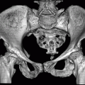
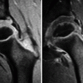
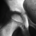
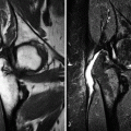
In acute avulsion fractures, radiographs show a bone fragment with incompletely corticated borders in the soft tissues adjacent to the expected apophyseal site, with no need for additional investigation, even though magnetic resonance imaging (MRI) allows for a more precise and detailed assessment (Fig. 10.1). Radiographs are often normal in patients with early-stage apophysitis, while those with chronic apophysitis often exhibit apophyseal fragmentation and enlargement, physeal widening, and periosteal reaction (Fig. 10.2), which are also well demonstrated on computed tomography (CT) (Fig. 10.3). It is important to have in mind that irregularity and fragmentation may also be found in normal apophyses as a developmental variant, which is deprived of significance if there are no clinical manifestations. On ultrasound (US), chronically inflamed apophyses usually present increased size and heterogeneous vascularity; there may be swelling of the adjacent soft tissues as well, and the tendon may appear thickened, hypervascular, and hyperechogenic. Magnetic resonance imaging (MRI) is the most sensitive and specific method for evaluation of chronic apophysitis, showing an edematous apophysis of increased size that often presents post-gadolinium enhancement; edematous changes are also found in the adjacent bone marrow, in the physis, and in the surrounding soft tissues (Figs. 10.4 and 10.5). Physeal widening may be subtle or marked, and even apophyseal avulsion is found in severe cases (Figs. 10.4, 10.6, and 10.7). Chronic apophysitis may present a misleading appearance on imaging and simulate an aggressive process, with mass effect, marked periosteal reaction and exuberant bone neoformation, in addition to significant bone marrow edema and soft-tissue swelling (Fig. 10.4). Correlation between clinical data and imaging findings is crucial in order to avoid unnecessary biopsies, as that the histopathological picture may lead to an erroneous diagnosis of sarcoma.
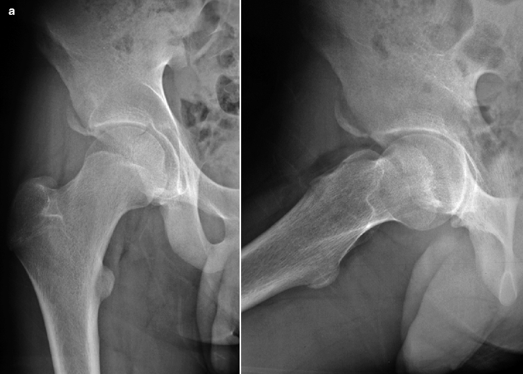
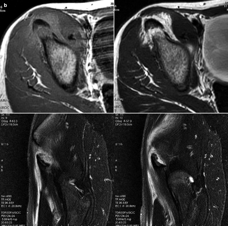
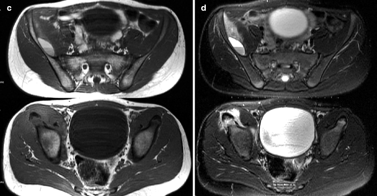
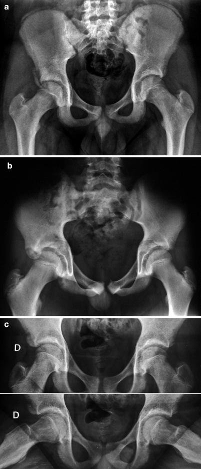
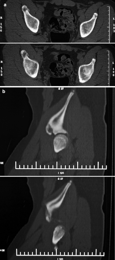
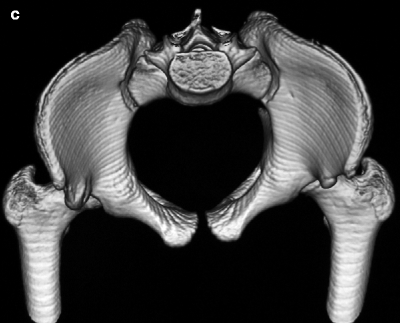
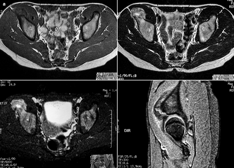
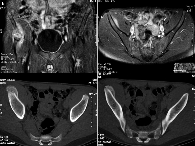
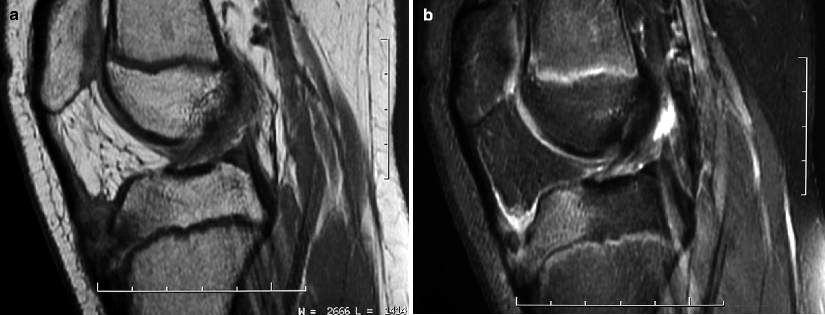
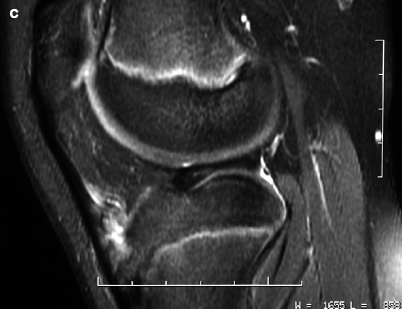
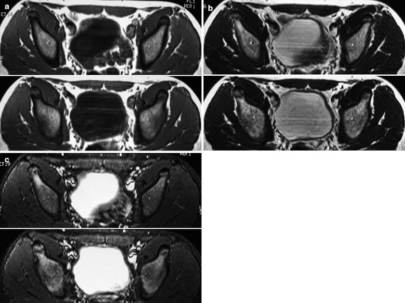
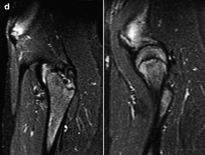
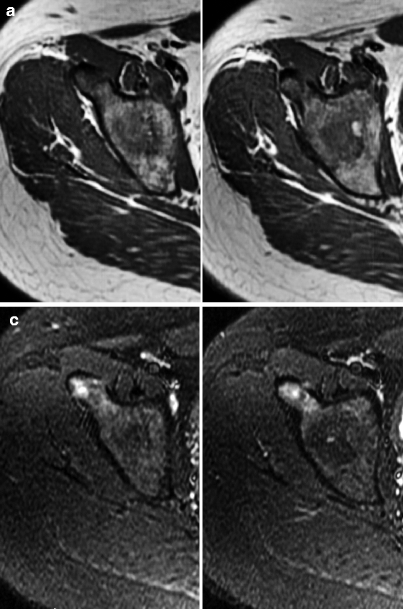
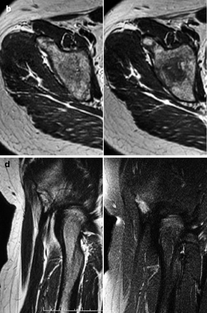


Fig. 10.1
In (a), radiographs of the right hip of an adolescent with acute avulsion of the anterior inferior iliac spine show a partially corticated bone fragment in the soft tissues adjacent to the apophyseal site, without evidence of chronic apophysitis. In (b), MRI of another patient demonstrates similar findings, with acute avulsion of the same apophysis and mild displacement, without signs of chronic inflammation. T1-WI (upper row, left) and T2-WI (upper row, right) demonstrate increased signal intensity of the periapophyseal soft tissues, related to posttraumatic hemorrhage, while sagittal fat sat T2-WI (lower row) reveal extensive soft-tissue edema and allow for an accurate estimate of the extent of the avulsion. Transverse T1-WI (c) and fat sat T2-WI (d) of the pelvis of the same patient disclose a large hematoma deeply situated relative to the right iliac muscle, containing a fluid-fluid level (Fig. 10.1a; courtesy of Dr. Marcelo Coelho Avelino, Medimagem, Teresina, Brazil)

Fig. 10.2
Radiographs of three distinct patients with right-sided chronic apophysitis of the anterior inferior iliac spine displaying different appearances of this condition on imaging. All of them evidence increased size and abnormal shape of this apophysis. In (a), periosteal reaction and bone neoformation are evident, which are notably absent in the acute lesion of Fig. 10.1a. In (b), the apophysis has a pseudotumoral appearance, with bone deformity, metaphyseal remodeling, and physeal widening. In (c), despite the obvious increase of the apophyseal size, bone neoformation/remodeling is absent


Fig. 10.3
CT of the pelvis of a 14-year-old soccer player. Transverse images (a), sagittal reformatted images (b), and volume-rendered reconstruction (c) display increased size of the apophysis of the right anterior inferior iliac spine, with physeal widening and bone neoformation. Apophysitis is more evident when the affected apophysis is compared to the contralateral one


Fig. 10.4
In (a), transverse T1-WI (upper-left image), T2-WI (upper-right image), and STIR image (lower-left image) reveal findings similar to those of the patient of Fig. 10.3; MRI, however, is able to detect edematous changes of the bone marrow and of the soft tissues, which are related to chronic inflammation and not evident on CT. Sagittal gradient-echo image (lower-right image) is useful to demonstrate increased size and irregular contours of the affected apophysis, as well as physeal widening. In (b), post-gadolinium fat sat T1-WI (upper row) display enhancement of the above-mentioned edematous areas, while CT images (lower row) evidence solid and thick periosteal neoformation, which is difficult to demonstrate on MRI


Fig. 10.5
Sagittal T1-WI (a), fat sat PD-WI (b), and post-gadolinium fat sat T1-WI (c) of a patient with apophysitis of the tibial tubercle show bone fragmentation in addition to thickening and heterogeneity of the distal portion of the patellar tendon. In addition, there is fluid distension of the deep infrapatellar bursa and edematous changes of the bone marrow and of the adjacent soft tissues, which show enhancement on post-contrast images


Fig. 10.6
MRI of the hips of a 14-year-old adolescent with chronic apophysitis of the right anterior inferior iliac spine. Transverse T1-WI (a), T2-WI (b), and fat sat T2-WI (c) reveal subtle increase of the size of the affected apophysis and adjacent bone marrow edema, which are absent in the opposite side. In (d), sagittal fat sat T2-WI demonstrate widening and increased signal intensity of the right physis, in addition to the above-mentioned findings


Fig. 10.7
A 15-year-old patient with chronic inguinal pain at right due to chronic apophysitis of the anterior inferior iliac spine. Transverse T1-WI (a), T2-WI (b), and STIR images (c) and sagittal T2-WI and fat sat T2-WI (d) display findings very similar to those of the patient of Fig. 10.6, unless for absence of significant physeal widening
Several apophyses may be affected in the pelvis and in the hips, such as the iliac crest, the anterior superior and anterior inferior iliac spines, the ischial tuberosity, and the greater trochanter. Apophysitis of anterior inferior iliac spine is more prevalent in countries where soccer is more popular, resulting from excessive traction exerted by the tendon of the direct head of the rectus femoris. Apophysitis around the hip typically does not affect individuals under age 13, given that the apophyseal ossification centers usually appear between 13 and 15 years of age and fuse between 16 and 18. Inclusion of both hips in the field of view is recommended for CT and MRI coronal and axial images, as comparative analysis of the apophyses maximizes the diagnostic capabilities of these methods (Figs. 10.1, 10.2, 10.3, 10.4, 10.6, and 10.7). Old healed lesions frequently lead to deformity of the affected apophysis, which displays density/signal intensity similar to those of the normal bone (Fig. 10.8).
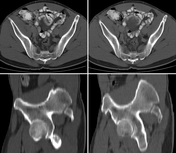

Fig. 10.8
Bone deformity related to an old healed lesion of the left anterior superior iliac spine, incidentally found during an abdominal CT scan of a 36-year-old male (former soccer player during the adolescence). On transverse images (upper row), there is prominence of the apophysis, with a bony protuberance that is also evident on a sagittal reformatted image (right lower image – compare with the normal apophysis in the left lower image)
Osgood-Schlatter disease (OSD) is a traction apophysitis of the insertion of the patellar tendon in the tibial tubercle. It is more frequent in male athletes between 10 and 15 years of age, occurring bilaterally in 20–30 % of the cases. In addition to the above-mentioned classic radiographic findings (Fig. 10.9), heterotopic intratendinous ossification is also common, as well as fluid distension of the deep infrapatellar bursa (Figs. 10.5, 10.10, and 10.11). Residual bone deformity and/or persistence of an intratendinous ossicle in the distal portion of the patellar tendon can be found after healing and may cause chronic pain (Fig. 10.12).
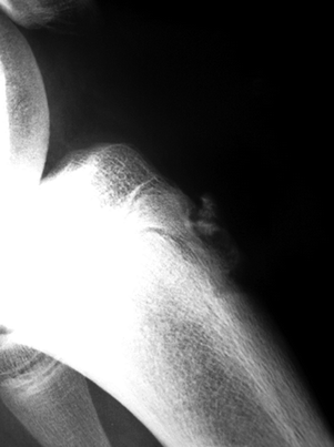
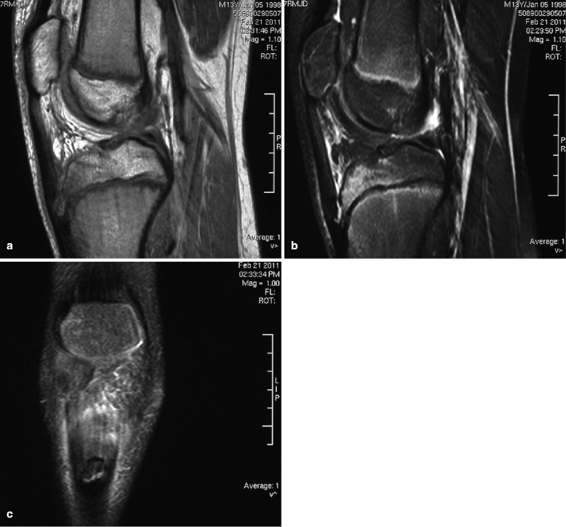
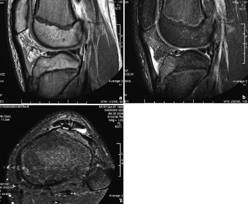
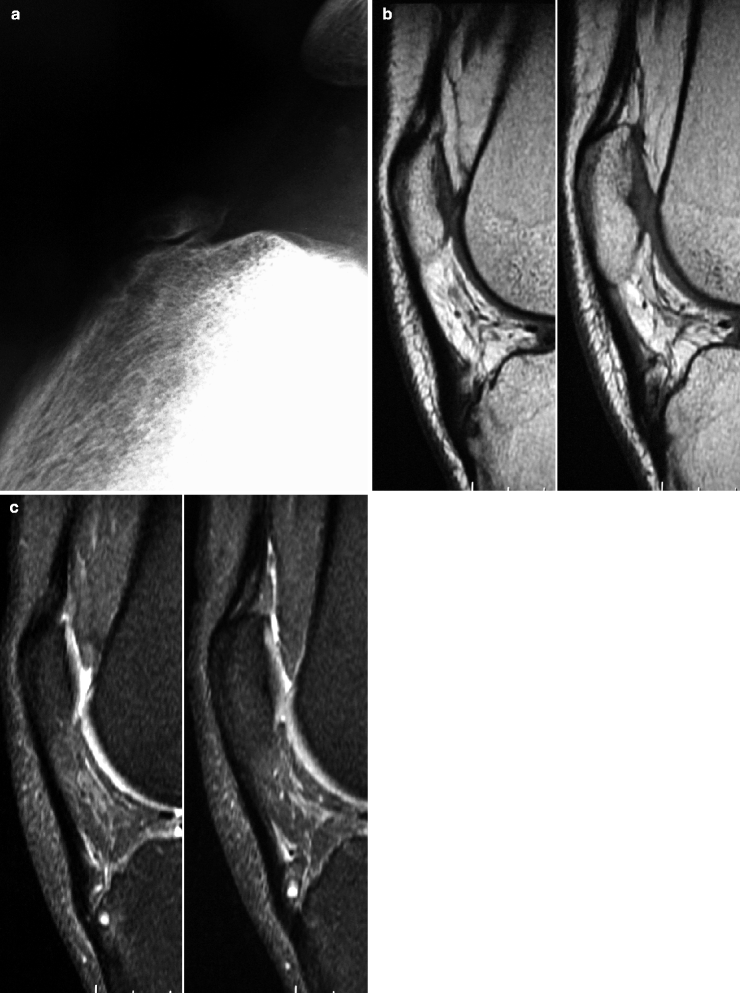

Fig. 10.9
Lateral view of the left knee of a child with OSD showing fragmentation and sclerosis of the tibial tubercle

Fig. 10.10
MRI of the right knee of a 13-year-old child with OSD. Sagittal T1-WI (a) and fat sat T2-WI (b) and coronal fat sat T2-WI (c) display fragmentation of the apophysis of the tibial tubercle and extensive bone marrow edema pattern, as well as edema of the infrapatellar fat pad and thickening of the distal portion of the patellar tendon

Fig. 10.11
A 13-year-old patient with OSD. In addition to the findings above described for the patient of Fig. 10.10, sagittal PD-WI (a) and STIR image (b) and transverse STIR image (c) disclose fluid in the deep infrapatellar bursa

Fig. 10.12
Lateral radiograph (a) and sagittal T1-WI (b) and fat sat PD-WI (c) of an adult with late-stage sequelae of OSD complaining of anterior knee pain. There is an ossicle projected over the distal portion of the patellar tendon, with irregularity of the tibial tubercle. MRI corroborates the radiographic findings, demonstrating cystic/edematous changes of the bone marrow of the tibial tubercle and of the ossicle, as well as thickening of the distal patellar tendon and edema of the adjacent soft tissues
Sinding-Larsen-Johansson disease (SLJD) is a reaction to traction at the inferior pole of the patella, more common in males between 9 and 12 years old, mainly those involved in jumping, gymnastics, and kicking sports. Strictly speaking, the inferior patellar pole is not an apophysis; nonetheless, the etiology and the inflammatory response to traction are identical to those seen in classic apophysitis. Imaging findings include bone fragmentation and inflammation in the inferior pole of the patella, occasionally associated with ossification of the proximal portion of the patellar tendon (Figs. 10.13, 10.14, and 10.15). Tendinopathy/partial tear of the patellar tendon (Fig. 10.16) and patellar fractures are the main differential diagnosis.
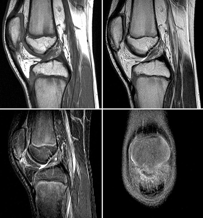
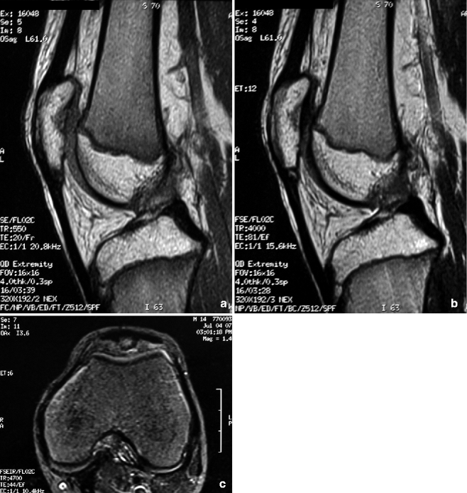
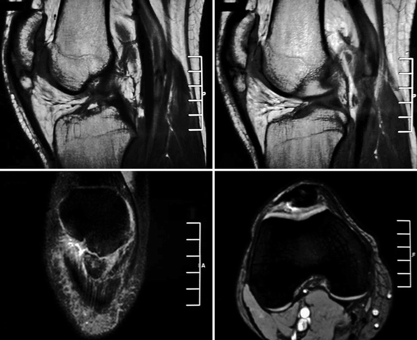
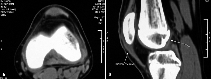

Fig. 10.13
MRI of the left knee of an 11-year-old soccer player with SLJD. Sagittal T1-WI (upper-left image), T2-WI (upper-right image), and STIR image (lower-left image) and coronal fat sat PD-WI (lower-right image) display bone marrow edema and fragmentation of the inferior pole of the patella; edema is also seen in the infrapatellar fat pad

Fig. 10.14
Sagittal T1-WI (a) and T2-WI (b) and transverse STIR image (c) of a 14-year-old patient with SLJD display bone fragmentation, more evident than that observed in the patient of Fig. 10.13. Nonetheless, the edematous changes are less prominent and almost exclusively confined to the avulsed bone fragment. Subtle thickening of the proximal patellar tendon is also present

Fig. 10.15
A 27-year-old patient with symptomatic sequelae of SLJD. MRI discloses chronic avulsion of a large bone fragment with corticated borders adjacent to the inferior patellar pole, as well as thickening of the proximal patellar tendon and edema of the adjacent soft tissues

Fig. 10.16
Transverse CT image (a) and sagittal reformatted image (b) of a 13-year-old runner with left infrapatellar pain disclose thickening and focal hypodensity of the proximal patellar tendon due to tendinopathy, without abnormalities of the adjacent bone or of the soft tissues
Sever’s disease is the inflammation of the calcaneal apophysis, adjacent to the insertion of the Achilles tendon. It is more common in males with less than 11 years of age, occurring bilaterally in up to 60 % of the patients. Pain worsens with physical activity and can be reproduced upon latero-lateral compression of the heel. As stated above, even though the classic findings of apophyseal fragmentation and sclerosis are present, the same findings may be found in normal individuals; additionally, recent works indicate that radiographs are not necessary in the workup as they have no real influence in the diagnosis or in therapeutic decision making. Similarly, bone marrow edema pattern may be found in the calcaneal apophysis of symptomatic and asymptomatic children on MRI. Therefore, there is no characteristic imaging pattern for this disease and the diagnosis is primarily a clinical one.
Stay updated, free articles. Join our Telegram channel

Full access? Get Clinical Tree


