Fig. 16.1
Relationship between method of PET scanning and image quality and their possible effects on diagnostic capability. NEC noise equivalent count, PVE partial volume effect
16.3.2 Injection Activity and Scan Time
Each PET camera model has its own intrinsic sensitivity and resolution. While newer models in general have better intrinsic performance, old cameras are still in use. A PET camera of lower intrinsic sensitivity needs larger injection activity and/or longer scan time to collect sufficient gamma-ray counts to generate images of sufficiently low statistical noise. It should be noted, however, that a limitation exists to the injected activity due to radiation exposure on the subject. Too much injected activity also degrades the image because higher count rate increases random counts and count loss and would only increase noise. Noise equivalent count (NEC), which is the count corrected for random and scatter, is often used instead of crude count to assess “effective” count.
16.3.3 Reconstruction Parameters
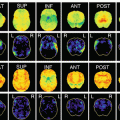
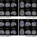
The reconstruction parameters, which are the parameters used for generating PET images in the computer after the subject is scanned, together with the post-filter, determine the trade-off between the image resolution and image noise. Iterative reconstruction algorithm called ordered subset expectation maximization (OSEM) and its modifications are often used for the reconstruction process, in which the number of subsets and the number of iterations are the two parameters to be determined by the investigator. By changing the reconstruction parameters, images of various resolution and noise are generated from the identical acquired data (Fig. 16.2). In principle, larger number of subsets or larger number of iterations generates an image of higher resolution but with larger noise.

Fig. 16.2
Effect of reconstruction parameters (number of iterations and subsets in OSEM) applied to an identical data acquired with a Discovery-690 PET/CT camera upon the FDG-PET images of a normal subject
16.3.4 Harmonization Between Cameras
In a multicenter study using various PET camera models, it is essential to determine appropriate scanning conditions for each PET camera model, especially scan time and reconstruction parameters, so that reliable images of similar quality could be obtained across different imaging sites. This process is called “harmonization” between different PET cameras, and phantoms are used for this purpose. In a well quality-controlled multicenter clinical research, the PET QC manager (PET QC Core) selects the phantom(s) and examines each PET camera to test the performance and to determine the appropriate scanning conditions before the PET camera is used for subject scans of the project.
16.4 Essential Image Quality for Brain PET
16.4.1 Key Image Quality to FDG-PET
FDG, a marker of glucose metabolism, accumulates high in the cerebral cortex and low in the white matter, creating a clear contrast between gray and white, which provides structural information on the PET images and helps identification of lesion localization. The area of high FDG uptake on the surface of the brain presents itself as a ring on the axial tomogram and is called cortical rim. From the standpoint of histology, the cortical rim is actually a mixture of gray and white matter tissues interlacing each other. Because of partial volume effect, the apparent FDG uptake in the cortical rim reflects the fraction of gray matter tissue more than the FDG uptake per gray matter tissue volume. If pathological cortical atrophy occurs, the apparent FDG uptake in the cortical rim is decreased, sometimes making it difficult to distinguish pathological tissue hypometabolism from apparently decreased uptake due to atrophy.
Detection of localized hypometabolism is a key to interpretation of FDG-PET images in research and diagnosis on dementia, because AD and some other neurodegenerative disorders each present a characteristic pattern of hypometabolism [1]. Reduced FDG uptake in the temporoparietal cortex and in the posterior cingulate and precuneus area is called AD pattern, which is useful for early or differential diagnosis of AD. To detect mild hypometabolism, PET images should have sufficient resolution and contrast together with sufficiently low noise. Furthermore, uniformity is also important because regional FDG uptake is evaluated in comparison with other areas.
In addition to visual interpretation, so-called statistical image analysis such as 3D-SSP is often used, in which the subject’s brain is spatially normalized to the standard coordinates system and the relative regional uptake is compared voxel by voxel with the normal database defined on the standard coordinates system to delineate areas of significant hypometabolism.
16.4.2 Key Image Quality to Amyloid-PET
In amyloid PET imaging, radioactivity uptake is observed in the cerebral cortex of subjects who have pathological deposition of amyloid beta plaque in corresponding areas. Positive scan is characterized by such abnormal uptake and is found in most AD patients and in some cognitively normal old subjects, while negative scan is characterized by the absence of such abnormal cortical uptake [2].
There are a number of PET drugs used for amyloid PET imaging, but all of them, more or less, accumulate nonspecifically in the white matter. Therefore it is necessary to detect mild cortical uptake adjacent to the nonspecific uptake by the white matter, which may often be a challenge, especially in subjects with cortical atrophy. Therefore, PET images again should have sufficient resolution and contrast together with sufficiently low noise. The contrast between gray and white evaluated in the phantom image is especially important to detect mild cortical uptake on the subject images.
In the quantitative analysis of amyloid PET images, which is used as an adjunct to the visual interpretation as well as for evaluation of the effect of therapeutics, the ratio of cortex to cerebellum or pons as a reference region, called standardized uptake value ratio (SUVR), is used. Therefore, uniformity within the field of view is also essential in amyloid PET.
16.5 Phantom Test Criteria for FDG and Amyloid-PET
16.5.1 Phantom Tests in J-ADNI
A large-scale multicenter study named J-ADNI, followed by a similar study J-ADNI2, was launched in Japan, in which subjects with normal cognitive function, mild cognitive impairment (MCI), and mild AD were to be followed with various examinations including FDG and amyloid PET scans. To standardize the PET scanning protocol across the participating PET centers and to harmonize the PET image quality between different PET cameras, the J-ADNI PET QC Core determined the subject preparation condition, standard injection activity, and accumulation time and established the phantom test procedures and criteria, with which to determine the reconstruction parameters for each PET camera.
In the camera qualification and harmonization procedures, two types of phantoms, i.e., Hoffman 3D brain phantom (Data Spectrum Corporation) and uniform cylindrical phantom, are used to measure a total of four image quality parameters: (1) resolution, (2) gray-white contrast, (3) uniformity, and (4) image noise. These four parameters have been adopted because they are considered essential for qualification of PET cameras and for harmonization of the PET image quality between PET cameras in multicenter clinical research on dementia using FDG and amyloid-PET as discussed above.
These four parameters have also been adopted in the phantom test criteria by the Japanese Society of Nuclear Medicine (JSNM) and published in the JSNM guidelines [3].
16.5.2 JSNM Phantom Test Criteria
The JSNM phantom tests prompt measurement of the four physical parameters (resolution, gray-white contrast, uniformity, and image noise) on the phantom images as discussed above.
Related posts:

Stay updated, free articles. Join our Telegram channel

Full access? Get Clinical Tree


