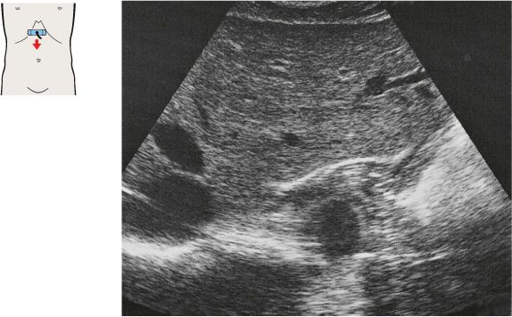
215 Esophagus, aorta, liver
216 Cardia, aorta, liver
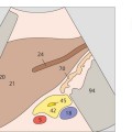
The gastroesophageal junction is identified between the liver, aorta, and crura of the diaphragm.
The cardia opens into a sharply tapered triangular structure when viewed in transverse section.
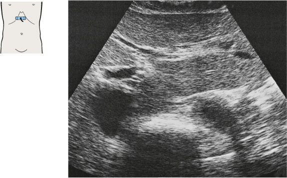
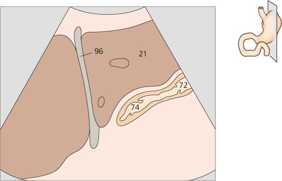
217 Cardia, gastric body, aorta, liver
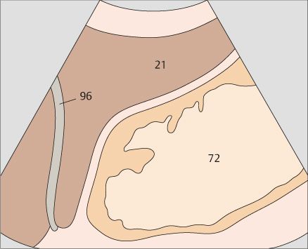
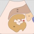
218 Gastric body, aorta, liver
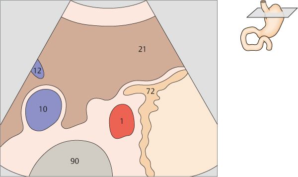
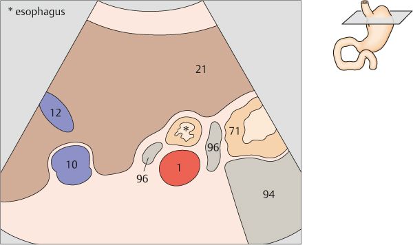
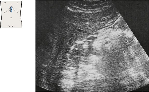
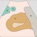
Next to the cardia, the gastric body presents a seemingly chaotic pattern of solid, liquid and gaseous material.
Below the cardia, the gastric body borders directly on the aorta.
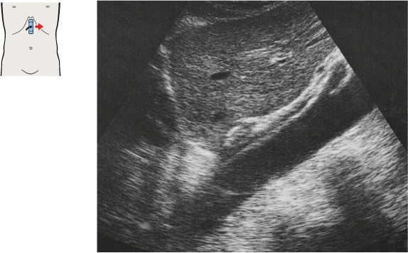
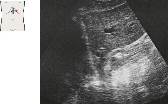
219 Esophagus, aorta, liver
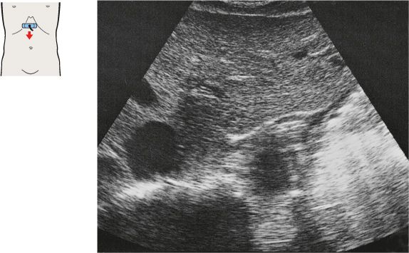
220 Esophagus, aorta, liver
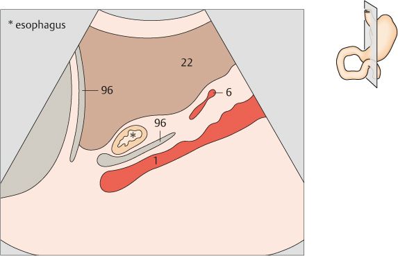
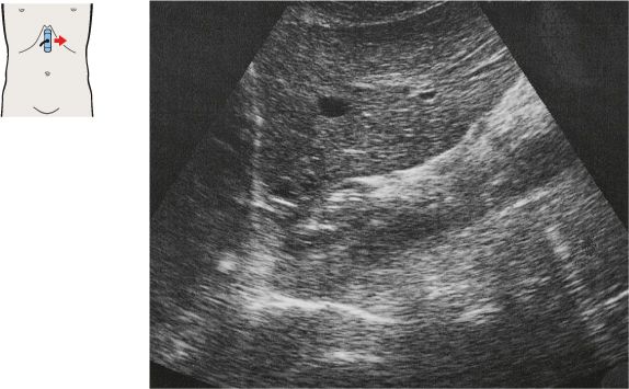
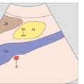
The abdominal esophagus is identified just to the right of, and anterior to, the aorta.
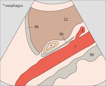
The esophagus and cardia are located between the liver and aorta in an upper abdominal longitudinal scan.
221 Cardia, liver
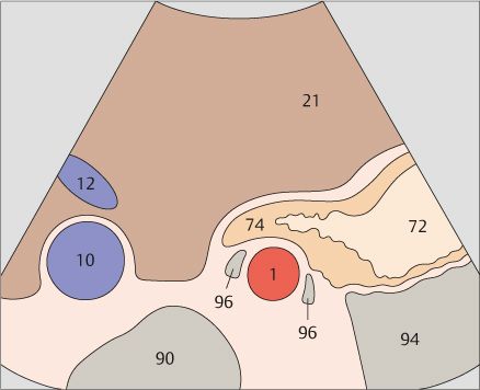
222 Gastric body, liver
The cardia and body of the stomach are located and identified by first defining the gastroesophageal junction.
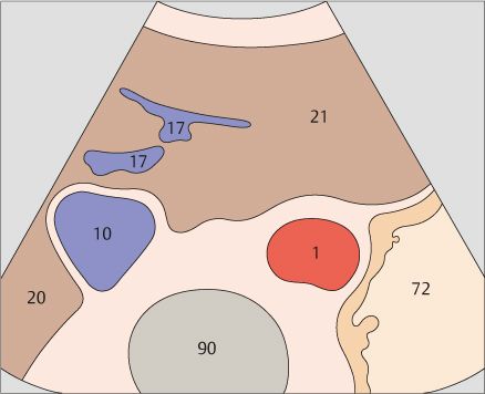
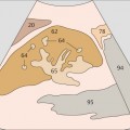
In subjects who have not been specially prepared, the gastric body presents a heterogeneous echo pattern located behind the left lobe of the liver.
Stay updated, free articles. Join our Telegram channel

Full access? Get Clinical Tree


